Mitochondrial peptides are small, mitochondrially encoded signaling microproteins, called MDPs, that include humanin, MOTS-c, and the SHLP family, alongside mitochondria-targeted synthetic peptides like SS-31 (elamipretide). They regulate cellular bioenergetics through retrograde signaling, AMPK activation, and cardiolipin stabilization. Preclinical evidence is strong and often dramatic; human clinical validation remains thin, constrained by bioavailability, stability, and delivery hurdles that any researcher working in this space needs to understand before designing a study.
TL;DR:
- Humanin, MOTS-c, SHLPs, and SS-31 vary in mechanism, with endogenous peptides acting as retrograde signals influencing gene expression and synthetic peptides targeting specific mitochondrial structures.
- SS-31 uniquely binds cardiolipin to prevent oxidation, maintaining electron transport chain integrity, and shows rapid bioenergetic improvements in aged mice.
- Human data is limited to associations, showing circulating MDP levels decline with age and rise with exercise, but causation and optimal dosing in humans remain unestablished.
- Developing mitochondrial peptides as drugs challenges stability and delivery, often requiring chemical modifications like D-amino acids, cyclization, or PEGylation to extend half-life.
- Future research needs standardized assays, human pharmacokinetic studies, and expanded characterization of new candidate peptides emerging from peptidomics efforts.
Synthrolab
Support Your Mitochondrial Peptide Research
Synthro Lab provides research-grade peptides and laboratory compounds for investigating mitochondrial function and related cellular signalling mechanisms.
Explore Synthro Lab
Table of Contents
What Are Mitochondrial Peptides? Origins and Classification
Mitochondrial DNA has traditionally been treated as a set of 37 genes coding for proteins used in oxidative phosphorylation and the RNA machinery that supports it. That picture turned out to be incomplete. Small open reading frames, or sORFs, embedded within the 12S and 16S mitochondrial rRNA genes encode a separate class of bioactive microproteins now called mitochondrial-derived peptides, or MDPs. At least eight of these have been characterized so far, including humanin, MOTS-c, and SHLP1 through SHLP6.
That number is almost certainly a floor, not a ceiling. sORF reannotation projects and mass-spectrometry-based peptidomics keep turning up candidate microproteins that fit the profile of an MDP but haven’t yet been functionally validated, which tells you the field is still in its early cataloging phase rather than its mature mechanistic one.
It helps to separate two categories that get lumped together under “mitochondrial peptides” in casual conversation:
- Endogenous MDPs — peptides your own mitochondrial genome transcribes and translates, including humanin, MOTS-c, and the SHLPs, which then act locally or get released into circulation as hormone-like signals.
- Mitochondria-targeted synthetic peptides — compounds like SS-31, engineered outside a biological genome specifically to accumulate in the inner mitochondrial membrane and act on a defined molecular target.
The distinction matters mechanistically. Endogenous MDPs function as a retrograde signaling network, meaning mitochondria are not just downstream recipients of nuclear instructions but active senders of signals back to the nucleus and to other tissues entirely. Researchers at the USC Cohen Lab describe this as a two-way communication system where mitochondrial stress or metabolic state gets broadcast outward, influencing gene expression in cells that never directly sensed the original stressor. Synthetic mitochondria-targeted peptides, by contrast, are designed interventions aimed at correcting a specific defect, such as cardiolipin oxidation, rather than mimicking a natural signaling event.
Physiologically, the roles cluster around three themes: cytoprotection (blocking apoptosis under metabolic stress), metabolic signaling (nudging AMPK and insulin sensitivity pathways), and systemic communication (acting as circulating mitokines that shift with age and exercise). Those three threads reappear constantly once you get into the specific peptides.
Humanin, MOTS-c, SHLPs, and SS-31: How Each One Works
Each of the major mitochondrial peptides operates through a distinct molecular route, and lumping them together as “mitochondrial support peptides” obscures more than it reveals. Here’s what separates them at the receptor and pathway level.
Humanin: blocking apoptosis through STAT3 and ERK
Humanin was discovered in surviving neurons from Alzheimer’s disease brain tissue, which set the tone for its entire research trajectory. It binds several extracellular receptor complexes and activates downstream STAT3 and ERK1/2 signaling, both of which suppress pro-apoptotic proteins like BAX. That gives humanin a fairly consistent cytoprotective signature across neuronal and cardiac tissue models: cells exposed to ischemic or oxidative stress survive at higher rates when humanin signaling is active. Its cardioprotective effects follow a similar logic, limiting cell death after simulated ischemia-reperfusion injury in isolated cardiomyocyte studies.
MOTS-c: the exercise-mimetic AMPK activator
MOTS-c is a 16-amino-acid peptide encoded within the 12S rRNA region, and it behaves almost like a metabolic alarm system. Under energy stress, MOTS-c activates AMPK, the same master regulator that exercise and caloric restriction engage, which is why researchers frequently describe it as an exercise mimetic in cell and rodent models. What makes MOTS-c mechanistically distinctive is that it doesn’t stop at the cytoplasm. Under metabolic stress, it translocates into the nucleus in an AMPK-dependent process and directly modulates nuclear gene expression tied to antioxidant defense, engaging NRF2 and antioxidant response element signaling along the way. Few other peptides in this category have a demonstrated nuclear-signaling arm, which is part of why MOTS-c gets so much attention in aging and insulin-resistance research.
SHLP1 through SHLP6: a family with opposite jobs
The Small Humanin-Like Peptides, numbered 1 through 6, share a genomic neighborhood with humanin but don’t share a single function. SHLP2 and SHLP3 tend toward protective, pro-survival signaling that overlaps with humanin’s cytoprotective role, supporting cell viability under metabolic stress and showing some influence over insulin secretion in beta-cell models. SHLP6 does close to the opposite: preclinical work associates it with pro-apoptotic signaling rather than protection. That split within a single peptide family is a useful reminder that “mitochondrial peptide” is not a synonym for “protective peptide.” Direction of effect depends entirely on which specific member you’re studying.
SS-31 (elamipretide): cardiolipin binding instead of ROS mopping
SS-31 works on a completely different principle than the endogenous MDPs above. It’s a synthetic mitochondria-targeted tetrapeptide that binds cardiolipin, the signature phospholipid of the inner mitochondrial membrane that organizes the electron transport chain into functional supercomplexes. Cardiolipin oxidation destabilizes those supercomplexes, causing electron leakage and driving up reactive oxygen species production at the source. SS-31’s primary mechanism is preventing that oxidation in the first place, preserving supercomplex assembly, rather than scavenging ROS after the fact once damage has already occurred.

That distinction, prevention versus cleanup, explains a pharmacologic property that sets SS-31 apart from most mitochondria-targeted antioxidants: it concentrates more than 1,000-fold in the inner mitochondrial membrane without relying on the mitochondrial membrane potential to get there. Compounds like TPP±conjugated antioxidants depend on an intact electrochemical gradient for uptake, which is precisely the thing that collapses in damaged or aging mitochondria. SS-31 sidesteps that problem entirely, which is why it retains activity even in severely compromised tissue where potential-dependent compounds lose their grip.
Pro Tip: When comparing peptide mechanisms in a grant proposal or literature review, specify whether you’re discussing a genomically encoded MDP or a synthetic mitochondria-targeted peptide. Reviewers increasingly expect that distinction up front, and blurring it is one of the more common critiques in peer review of this literature.
Across all four peptide families, one convergence point keeps showing up: cross-talk with PGC-1α and mitophagy regulation. MOTS-c and humanin both intersect with pathways that govern mitochondrial biogenesis and quality control, suggesting that MDPs aren’t just reactive stress signals. They may function as part of the same regulatory loop that decides whether a stressed mitochondrion gets repaired, replaced, or cleared.
What the Preclinical and Clinical Evidence Actually Shows
Preclinical data for mitochondrial peptides is genuinely striking in places. Human data is thin, inconsistent in methodology, and years behind the mechanistic story. That gap is the single most important thing to internalize before designing any translational study in this space.
The most cited rodent result involves SS-31 in aged mice. A single treatment restored mitochondrial energetics within one hour, reversing age-related declines in ATP production and mitochondrial coupling efficiency, and repeated dosing improved skeletal muscle endurance. A one-hour rescue effect in aged tissue is not a subtle finding. It’s the kind of result that makes SS-31 a recurring reference point in mitochondrial pharmacology, precisely because so few interventions produce measurable bioenergetic recovery on that timescale.
MOTS-c has its own preclinical track record, primarily around metabolic markers and exercise capacity in rodent models, consistent with its role as an AMPK-driven metabolic regulator. The mechanistic story (nuclear translocation, NRF2 engagement, insulin-sensitivity effects) lines up with observed improvements in glucose handling and exercise performance in animal work.
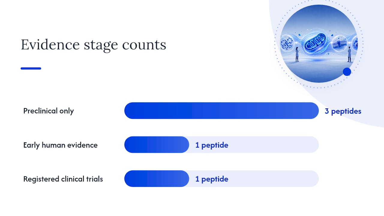
Human data tells a different kind of story, mostly observational rather than interventional. Circulating MDP levels decline with age and rise in response to acute and chronic exercise, a pattern consistent across several cohort studies. Low circulating MDP levels also associate with metabolic disease markers, though association is not causation, and most of these studies can’t establish which direction the relationship runs.
On the interventional side, elamipretide has the most developed clinical trial footprint of any peptide in this category, with trials registered on ClinicalTrials.gov covering conditions like primary mitochondrial myopathy and heart failure with preserved ejection fraction. Endpoints in these trials typically track functional measures such as six-minute walk distance and biomarkers of mitochondrial function rather than mortality outcomes, and trial durations tend to run in the weeks-to-months range rather than years. Humanin and MOTS-c, by comparison, remain almost entirely in preclinical and early translational research, without the same depth of registered human trial activity.
| Peptide |
Primary evidence stage |
Representative model or trial context |
| Humanin |
Preclinical (cell, some rodent) |
Neuronal and cardiomyocyte ischemia models |
| MOTS-c |
Preclinical, early human observational |
Rodent metabolic/exercise models; human circulating-level studies |
| SHLP2/SHLP3 |
Preclinical |
Beta-cell and cytoprotection models |
| SHLP6 |
Preclinical |
Pro-apoptotic signaling models |
| SS-31 (elamipretide) |
Preclinical plus registered clinical trials |
Aged mouse energetics; myopathy and heart failure trials |
The translational bottleneck isn’t a mystery. It comes down to pharmacokinetics and pharmacodynamics that remain poorly characterized in humans, combined with small sample sizes in the few interventional trials that exist. A rodent study with a dozen animals per group can detect a large effect size; a human trial needs either a much larger cohort or a much larger effect to reach the same statistical confidence, and mitochondrial peptide trials to date have leaned on the smaller end of both.
- Rodent and cell-model evidence for SS-31 and MOTS-c is consistently strong across independent labs.
- Human observational data supports age and exercise associations but can’t establish mechanism or causation in people.
- Registered clinical trials exist primarily for SS-31, with humanin and MOTS-c still largely confined to preclinical work.
- PK/PD characterization in humans lags well behind the mechanistic understanding built in animal models.
Measuring Mitochondrial Peptides: Assays and Lab Handling
Getting a reliable read on MDP levels or peptide activity in a research setting depends on choosing the right assay and respecting the pre-analytical variables that can quietly wreck a dataset.
- Choose the assay based on your question. ELISA-based methods are faster and cheaper for tracking relative changes in humanin, MOTS-c, SHLP2, or SHLP6 across a treatment timeline, but antibody specificity varies and cross-reactivity between SHLP family members is a documented concern. Mass-spectrometry approaches, particularly targeted LC-MS/MS, offer better specificity and can distinguish closely related peptide sequences, at the cost of more expensive instrumentation and longer sample turnaround.
- Standardize pre-analytical handling before you standardize anything else. Sample type (plasma versus serum), anticoagulant choice, and time-to-freeze all affect measured peptide concentrations, and inconsistent handling across a study is one of the more common sources of unexplained variance in MDP research.
- Store and freeze consistently. Most peptide assays call for rapid freezing at negative 80 degrees Celsius and minimizing freeze-thaw cycles, since repeated thawing degrades peptide integrity and skews concentration readings downward over time.
- Build in spike-recovery controls. Running a known concentration spike alongside your samples tells you whether your matrix is interfering with detection, which matters more with plasma than with simpler buffer systems.
- Report your limit of detection and inter-assay coefficient of variation. Reviewers increasingly expect these numbers alongside raw concentration data, since two labs reporting different absolute values for the same peptide are often just reporting different assay sensitivities, not different biology.
Pro Tip: If you’re running a longitudinal study tracking MDP changes over weeks or months, run all timepoints on the same assay plate or the same LC-MS/MS batch whenever possible. Batch-to-batch assay drift is a real and underappreciated source of false trend detection in this literature.
For synthetic peptides like SS-31 used in your own bench work, a certificate of analysis confirming purity and identity by HPLC or mass spec, combined with endotoxin testing, should be a baseline requirement before the compound goes anywhere near a cell culture plate or animal protocol. Batch purity issues are a quiet but common confound in peptide signaling research, since a contaminated or degraded batch can produce an apparent effect (or a null result) that has nothing to do with the peptide’s actual biology.
Why Mitochondrial Peptides Are Hard to Develop as Drugs
Short peptides face a predictable set of liabilities, and mitochondrial peptides are no exception. Proteolytic degradation by circulating and tissue peptidases can chew through an unmodified peptide in minutes. Renal filtration clears small molecules quickly given their low molecular weight. The combined result is often a half-life measured in single-digit minutes for an unmodified sequence, which is workable for a cell-culture experiment and largely impractical for a human dosing regimen without either very frequent administration or chemical modification.
Several stabilization strategies address these liabilities directly:
- D-amino acid substitution replaces vulnerable L-amino acids at key positions, since most proteolytic enzymes are stereospecific and can’t efficiently cleave the D-form.
- Cyclization locks the peptide into a constrained conformation that’s harder for exopeptidases to gain purchase on.
- PEGylation attaches polyethylene glycol chains that increase molecular size and reduce renal clearance, at the cost of altered tissue penetration.
- Lipid carrier conjugation improves membrane permeability and can extend circulation time, particularly relevant for peptides that need to reach an intracellular or intramitochondrial target.
SS-31 illustrates why mechanism-driven design matters more than simply chasing membrane potential. Instead of relying on the electrochemical gradient that potential-dependent delivery systems require, its alternating cationic and aromatic residues let it partition directly into the inner mitochondrial membrane and bind cardiolipin regardless of how compromised that gradient already is. That’s a fundamentally different design philosophy from earlier mitochondria-targeted antioxidants, and it’s part of why SS-31 has made it further into clinical testing than most peptides in this category.
None of this comes cheap. Peptide synthesis costs scale with sequence length, the number of non-standard amino acids, and the purity threshold required for a given application, and translational reviews consistently flag manufacturing cost as a real constraint on how far a promising mitochondrial peptide can progress through preclinical development before funding runs out.
A Researcher’s Checklist for Sourcing and Handling Mitochondrial Peptides
Before a mitochondrial peptide ever touches a plate or an animal, a handful of sourcing and handling decisions determine whether your downstream data will hold up to scrutiny.
- Verify the certificate of analysis before you order, not after. A legitimate COA states purity percentage (typically confirmed by HPLC), identity confirmation (usually mass spectrometry), and batch number. If a supplier can’t produce one on request, treat that as a disqualifying red flag rather than a minor inconvenience.
- Insist on independent purity verification for critical experiments. Supplier-reported purity is a starting point, not a guarantee; for high-stakes work, third-party verification of at least a sample lot adds a real layer of confidence.
- Check the acceptable impurity profile, not just the headline purity number. A peptide listed at 95% purity could have its remaining 5% distributed across many trace impurities or concentrated in one problematic byproduct, and that distinction matters for anything sensitive to off-target effects.
- Confirm endotoxin levels before cell culture or in vivo work. Endotoxin contamination can produce inflammatory signaling that’s easy to misattribute to the peptide itself if you haven’t tested for it directly.
- Match your reconstitution solvent to the peptide’s solubility profile. Getting this wrong is one of the most common avoidable errors in peptide handling, and it’s worth checking supplier-specific reconstitution guidance rather than assuming one solvent fits every sequence.
- Store lyophilized peptide at negative 20 degrees Celsius or lower, and aliquot reconstituted stock to avoid repeated freeze-thaw cycles. Peptide stability drops measurably with each freeze-thaw event, and aliquoting up front costs almost nothing compared to the cost of a compromised experiment.
- Keep every product clearly labeled “for laboratory research use only.” Mitochondrial peptides sold through research-grade suppliers, including Synthro Lab, are intended for laboratory investigation, not human administration, and any work intended for human subjects requires its own separate IRB or IND regulatory pathway entirely distinct from research-use sourcing.
Pro Tip: Keep a running log of lot numbers alongside your experimental data, not just in a separate inventory spreadsheet. If you see an unexplained shift in your results, the first thing to cross-check is whether it coincides with a new batch, and that log is what makes the check possible in five minutes instead of an afternoon.
Where the Research Needs to Go Next
The biggest open question in this field isn’t whether mitochondrial peptides do something interesting. Preclinical data has settled that. The real gap is human pharmacokinetics and pharmacodynamics, which remain sparse even for SS-31, the most clinically advanced compound in this category, and are close to absent for humanin and MOTS-c.
A second, quieter problem is receptor identification. Humanin’s receptor complexes are reasonably well described, but MOTS-c’s precise receptor mechanism is still incompletely mapped, which limits the ability to design targeted agonists or predict off-target effects with confidence.
A few study designs would move the field faster than incremental animal work:
- Short, well-controlled human PK/PD studies for MOTS-c and humanin analogs, even at small scale, to establish basic dosing and clearance parameters.
- Adaptive small-sample efficacy trials that use biomarker endpoints (mitochondrial function assays, exercise capacity) rather than waiting for large outcome trials that this early-stage field can’t yet justify.
- Multi-site assay standardization efforts, so that an MDP concentration reported by one lab means the same thing as a concentration reported by another.
- Public data sharing for peptidomics and sORF discovery work, since new MDP candidates are being identified faster than the field can currently validate them individually.
None of that requires a breakthrough. It requires the kind of methodological discipline that’s often less exciting to fund than a new mechanism paper, but without it, the gap between rodent data and human application will keep widening rather than closing.
Beyond Humanin and MOTS-c: What’s Coming Next in MDP Research
The eight characterized MDPs (humanin, MOTS-c, and SHLP1 through SHLP6) are very likely not the final list. Advances in sORF reannotation and mass-spectrometry-based peptidomics keep surfacing candidate microproteins encoded within mitochondrial DNA regions that were previously assumed to be non-coding or purely structural.
That expansion matters for two reasons. First, some of these newly flagged candidates may fill functional gaps the current MDP roster doesn’t cover well, particularly around tissue-specific signaling that humanin and MOTS-c don’t fully explain on their own. Second, and more practically for anyone running assays today, a growing candidate list means antibody-based detection methods will need continuous updating to avoid cross-reactivity with peptides that weren’t even known to exist when the assay was originally validated.
The near-term direction for the field looks less like discovering an entirely new signaling paradigm and more like filling in a peptide family tree that’s already partially sketched. Expect incremental additions to the known MDP list, better resolution of which peptides act through overlapping receptors versus distinct ones, and continued work on whether some of these emerging candidates share SS-31’s design logic of targeting a specific mitochondrial structure rather than acting as a broad-spectrum antioxidant.
An Editorial Take on Where This Field Actually Stands
The conventional pitch on mitochondrial peptides oversells the clinical readiness and undersells how genuinely interesting the mechanistic story is. That’s backwards. SS-31’s one-hour energetic rescue in aged mice and MOTS-c’s nuclear signaling arm are the kind of findings that should excite anyone doing bioenergetics research, not because they promise a therapy tomorrow, but because they reveal mitochondria as active signaling hubs rather than passive energy factories.
Where the field oversells itself is skipping straight from “works in a mouse” to implied human relevance, without dwelling on the PK/PD gap that separates the two. Where it undersells itself is treating this as a niche curiosity rather than a mechanism with implications across metabolic, cardiovascular, and neurodegenerative research. My honest read: prioritize the peptides with the clearest mechanistic story and the most rigorous COA and purity data behind them, run your own PK groundwork before assuming translation, and treat batch quality as a variable worth controlling as tightly as any other in your protocol.
— Mitch
Research-Grade Support for Mitochondrial Peptide Studies
Reliable mitochondrial peptide research starts with knowing exactly what’s in the vial. Research-grade peptides relevant to mitochondrial and metabolic signaling work are available from suppliers that provide independent batch testing and certificates of analysis, so purity and identity can be verified before experiments start.
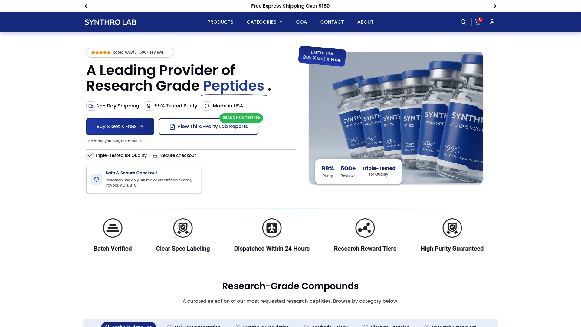
Beyond the peptides themselves, Synthrolab’s educational resource on peptide types and quality standards walks through what a proper COA should contain and how purity thresholds affect experimental reliability, useful groundwork whether you’re new to peptide sourcing or auditing an existing protocol. Researchers working on metabolic and bioenergetic pathways can also review Synthrolab’s guide to metabolic health compounds for related context on lab-relevant metabolic signaling. If your lab needs bulk quantities, specific stability documentation, or COA verification for a particular batch, consider contacting product suppliers directly to discuss research inquiries before placing an order.
Recommended
 21+ verification