At the cellular level, joint health is the homeostatic state in which articular cartilage, synovium, and subchondral bone maintain their structural integrity and functional capacity through balanced ECM synthesis and degradation, regulated immune signaling, and intact organelle function. The dominant mechanistic framework shaping research in 2026 is the ECM–mitochondria–mechanics axis: joint fate is determined by how extracellular matrix remodeling, organelle dysfunction, and mechanical loading converge with inflammatory signaling to tip tissue toward repair or degeneration.
For lab teams, the practical entry points are:
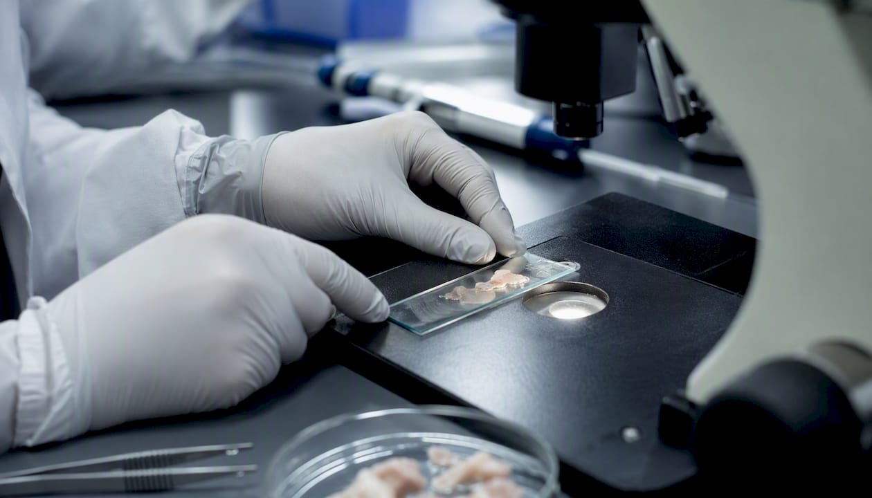
- Histological scoring: OARSI histopathology remains the gold-standard structural endpoint in preclinical models
- Transcriptomic resolution: Single-cell RNA-seq and spatial transcriptomics platforms, particularly 10x Genomics Visium, now resolve cell-state-specific drivers that bulk assays miss
- Peptide-based modulation: Molecules like p[63-82], RGD-family peptides, GHK-Cu, BPC-157, and TB-500 serve as mechanistic probes and candidate disease-modifying agents
Key Takeaways
Joint health at the cellular level is governed by the ECM–mitochondria–mechanics axis, and peptide-based interventions require multi-model validation, organelle-level readouts, and tight delivery controls to produce interpretable preclinical data.
| Point |
Details |
| Core mechanistic axis |
ECM–mitochondria–mechanics axis drives joint fate; measure ROS, mtDNA, and NLRP3 alongside structural endpoints. |
| Priority assays |
Run OARSI histology, SOX9/COL2A1 ratio, and IL-1β/TNF-α cytokine panel as the minimum interpretable endpoint set. |
| Peptide delivery design |
IA dosing requires weekly injections for unmodified peptides; cyclization improves stability but must be validated for retained bioactivity. |
| Model validation |
Positive results in MMT must be replicated in ACLT-pMMx before any translational claim; in vitro potency does not predict in vivo efficacy. |
| Synthrolab resources |
Batch-specific COAs and research-grade compounds (GHK-Cu, BPC-157, TB-500) support reproducible joint-health experiments. |
Table of Contents
What tissues and cell types define joint health in research?
Joint research spans four compartments, each with distinct resident cells and priority markers.
Articular cartilage is avascular and depends entirely on chondrocytes for ECM maintenance. These cells produce collagen II (COL2A1), aggrecan (ACAN), and the transcription factor SOX9, which together define the anabolic cartilage phenotype. Loss of SOX9 expression reliably precedes structural breakdown.
Synovium lines the joint cavity and regulates the biochemical environment through synoviocytes (fibroblast-like FLS and macrophage-like MLS). PRG4 (lubricin) production marks healthy FLS function; IL-6 and TNF-α secretion marks activated, pro-inflammatory states. The synovium is also the primary site of immune cell infiltration.
Subchondral bone couples mechanically and biochemically with cartilage via osteoblasts and osteoclasts. Remodeling here accelerates during OA progression and feeds back to cartilage through altered stiffness and cytokine gradients.
Meniscus contains fibrochondrocytes that produce both fibrocartilaginous (COL1A1) and hyaline-like (COL2A1) matrix. Meniscal integrity is a key variable in the MMT preclinical model.
Immune cell contributions are substantial. M1-polarized macrophages (CD68+/CCR2+) drive catabolic signaling; M2 phenotypes support resolution. Sample synovial fluid and synovial tissue separately to capture both resident and infiltrating populations.
| Tissue |
Dominant cell types |
High-priority research markers |
| Articular cartilage |
Chondrocytes |
COL2A1, ACAN, SOX9, MMP-13 |
| Synovium |
FLS, MLS, macrophages |
PRG4, IL-6, CD68, CCR2 |
| Subchondral bone |
Osteoblasts, osteoclasts |
RUNX2, OPG, RANKL |
| Meniscus |
Fibrochondrocytes |
COL1A1, COL2A1, OARSI score |
How does the ECM–mitochondria–mechanics axis govern joint fate?
Sustained inflammation combined with organelle dysfunction creates a feedforward loop: mitochondrial ROS production impairs chondrocyte energy metabolism, ER stress triggers unfolded protein responses, and lysosomal membrane permeabilization releases cathepsins that amplify ECM degradation. Mechanically, chondrocytes sense load through Piezo1/2 channels, TRPV4, ERα, and YAP, which convert loading into transcriptional programs affecting COL2A1 expression and hypertrophic gene activation.
Signaling pathway timing matters enormously. Stage-specific OA models show NF-κB and MAPK dominate early inflammation, AMPK suppression and mTOR activation characterize progressive disease, and Wnt/FGF upregulation marks late-stage catabolism. Designing interventions without accounting for this sequence is a common source of irreproducible results.
| Pathway |
Role in joint tissue |
Key readouts |
| TGF-β/BMP |
ECM synthesis, chondrogenesis |
SOX9, COL2A1, SMAD2/3 |
| Wnt/β-catenin |
Hypertrophy, catabolism |
MMP-13, RUNX2, β-catenin |
| NF-κB |
Inflammatory amplification |
IL-1β, TNF-α, COX-2 |
| MAPKs (ERK) |
Stress response, apoptosis |
p-ERK, caspase-3 |
| AMPK/mTOR |
Metabolic homeostasis |
p-AMPK, p-mTOR, autophagy flux |
| Hedgehog |
Subchondral remodeling |
IHH, PTHrP, COL10A1 |
Pro Tip: When stimulating BMP/TGF-β repair pathways, co-measure RUNX2 and COL10A1 from the first time point. Hypertrophic drift often appears before structural changes are visible, and catching it early lets you adjust dose or delivery schedule before the experiment is compromised. TGF-β/BMP signaling requires tight spatial and temporal control to avoid Wnt-driven MMP-13 upregulation.
Which experimental models and assays answer which mechanistic questions?
In vitro systems each answer a distinct question:
- Primary chondrocytes: cytokine response, gene expression, and short-term drug screening (24–72 hours)
- Micromass pellet chondrogenesis: matrix production capacity and SOX9-driven differentiation over 14–21 days
- Cartilage explants: ECM turnover under near-native conditions, GAG release assays, 7–28 days
- Organoids and organ-on-chip: multicellular crosstalk and mechanical loading responses, 14–28 days
In vitro chondrogenesis readouts do not guarantee in vivo efficacy. Validate across mechanistically distinct preclinical models before claiming disease-modifying potential.
| Model |
What it measures |
Timeline |
Primary endpoints |
| MMT (medial meniscal tear) |
Cartilage degeneration, pain |
4–8 weeks |
OARSI score, weightbearing, gait |
| ACLT-pMMx |
Rapid OA progression, ECM loss |
8 weeks |
OARSI score, COL2A1, behavioral assays |
| Micromass pellet |
Chondrogenic differentiation |
14–21 days |
GAG content, COL2A1, SOX9 |
| Cartilage explant |
ECM turnover, cytokine response |
7–28 days |
GAG release, MMP activity |
For histology, Safranin O/Fast Green staining with OARSI scoring is the standard. Pair it with immunohistochemistry for COL2A1 and MMP-13 to distinguish anabolic from catabolic states. Behavioral readouts in rodent models should include incapacitance testing and von Frey filament assays for pain sensitization.
What peptides and biologics probe or modulate joint mechanisms?
Chondroinductive peptides fall into three functional classes: growth factor-derived, adhesion motif-derived (RGD family), and ECM-targeting sequences. Each class engages distinct receptors and downstream programs.
- p[63-82] (BMP7-derived): Chondroprotective via SMAD/NKX3-2 signaling; demonstrated reduced OARSI scores in MMT and ACLT-pMMx rat models with weekly IA dosing. A primary DMOAD candidate in preclinical literature.
- RGD-family peptides: Engage integrin receptors to promote chondrocyte adhesion and matrix deposition; commonly functionalized onto hydrogel scaffolds for IA delivery.
- LPP (link protein N-terminal peptide) / LPP-like sequences: Stimulate aggrecan synthesis via CD44 receptor engagement; useful for probing proteoglycan restoration.
- PEDF 29-mer: Combined with hyaluronic acid, promotes BM-MSC chondrogenic differentiation and cartilage regeneration in MIA rat models via PEDFR/STAT3 signaling. SOX9, ACAN, and COL2A1 all increase in vitro.
- GHK-Cu: Copper-binding tripeptide with anti-inflammatory and ECM-remodeling properties; modulates MMP expression and supports collagen synthesis in connective tissue models.
- BPC-157: Pentadecapeptide with cytoprotective and angiogenic properties; studied in tendon and ligament repair models with relevance to periarticular tissue.
- TB-500 (thymosin beta-4 fragment): Promotes actin dynamics and cell migration; relevant to synovial repair and inflammatory resolution models.
Delivery route is a critical variable. IA injection concentrates peptide at the target site but requires frequent dosing (typically weekly in rodent models) due to rapid synovial clearance. Sustained-release carriers, including hyaluronic acid hydrogels and PLGA microspheres, extend residence time and reduce injection frequency.
Pro Tip: Cyclization and N-/C-terminal modifications (N-acetylation, C-amidation) improve protease resistance in the synovial environment, but each modification must be validated for retained bioactivity. A cyclized variant that survives 24 hours in synovial fluid but loses receptor affinity is not an improvement. Run parallel stability and activity assays before committing to a modified sequence. See growth factor peptide classes for modification context.
Combining anabolic and immunomodulatory strategies is emerging as a required design principle. Depleted regenerative stem cell populations and expanded inflammatory macrophage phenotypes represent dual targets, which single-mechanism peptides often fail to address simultaneously.
Which molecular endpoints matter most in joint-health studies?
Prioritize these readouts in the order listed when assay budget is constrained:
- MMP-13 and ADAMTS5: Primary catabolic markers; elevated levels confirm active ECM degradation
- COL2A1 and ACAN: Anabolic markers; their ratio to MMP-13 indicates net matrix balance
- SOX9: Master chondrogenic transcription factor; a SOX9/COL2A1 ratio shift downward signals phenotypic drift toward hypertrophy
- COL10A1 and RUNX2: Hypertrophy markers; measure from day 7 onward in any chondroinductive experiment
- IL-1β, IL-6, TNF-α: Cytokine panel for inflammatory state; sample synovial fluid and tissue separately
- ROS and mtDNA: Organelle stress markers; elevated cytoplasmic mtDNA indicates mitochondrial membrane compromise
- NLRP3 and lysosomal markers (LAMP1, cathepsin B): Inflammasome activation and lysosomal membrane permeabilization
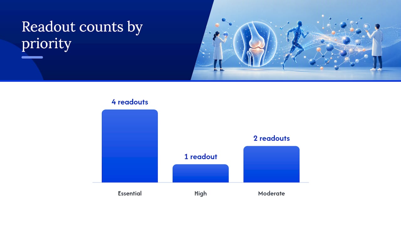
| Readout |
Priority |
Interpretation note |
| MMP-13 / ADAMTS5 |
Essential |
Elevated = active catabolism; normalize to tissue weight |
| COL2A1 / ACAN |
Essential |
Anabolic state; pair with SOX9 for phenotype confirmation |
| SOX9 |
Essential |
Downshift signals hypertrophic or catabolic drift |
| IL-1β / TNF-α |
Essential |
Confirm inflammatory context before interpreting ECM data |
| COL10A1 / RUNX2 |
High |
Hypertrophy screen; measure early, not just at endpoint |
| ROS / mtDNA |
Moderate |
Organelle dysfunction confirmation; useful in OA progression models |
| NLRP3 / LAMP1 |
Moderate |
Inflammasome and lysosomal pathway activation |
Regional heterogeneity is a real sampling problem. Medial vs. lateral cartilage zones differ in load history and cell density. Always specify the exact anatomical region sampled and keep it consistent across animals and time points.
How should you design your multiomics and imaging workflow?
A well-structured pipeline captures cell states, spatial context, and organelle function in parallel rather than sequentially.
- Single-cell RNA-seq: Dissociate tissue immediately post-harvest; use cold protease digestion to preserve chondrocyte viability. Cluster analysis should resolve chondrocyte subpopulations (homeostatic, pre-hypertrophic, effector) and synovial macrophage phenotypes (M1/M2 spectrum).
- Spatial transcriptomics (10x Genomics Visium): Cryosection at 10 µm; prioritize sections through the weight-bearing zone. Spatial deconvolution maps cell-state proportions to anatomical regions, revealing whether a peptide intervention shifts cell states in loaded vs. unloaded cartilage.
- Proteomics: Synovial fluid proteomics captures secreted MMP and cytokine profiles without tissue destruction; pair with tissue lysate proteomics for intracellular pathway confirmation.
- Imaging: Confocal microscopy for immunofluorescence co-localization (SOX9/COL2A1); second-harmonic generation (SHG) imaging for collagen fibril organization without staining artifacts.
Pro Tip: Preserve spatial integrity by processing loaded and unloaded tissue regions as separate samples from the start. Pooling them at extraction erases the mechanotransduction signal you are trying to detect. Pair mechanotransduction readouts (Piezo1, YAP localization) with spatial transcriptomics to map where mechanical inputs are actively shaping transcriptional programs.
Common analysis tools: Seurat and Scanpy for scRNA-seq clustering, CellChat or NicheNet for ligand-receptor inference, and STARmap or Squidpy for spatial deconvolution.
What experimental design rules make joint-health studies reproducible?
- Dose escalation: Test at least three dose levels per peptide; a single dose obscures the therapeutic window and makes safety assessment impossible.
- Dosing frequency: Match to synovial half-life. Weekly IA injections are standard for unmodified peptides in rodent models; sustained-release formulations may extend to biweekly.
- Route: IA delivery for joint-specific mechanistic questions; systemic delivery only when studying whole-body inflammatory modulation.
- Timing relative to injury: Early intervention (days 1–3 post-injury) tests prevention of degeneration; delayed intervention (weeks 2–4) tests repair capacity. Both are needed for translational relevance.
- Spatial targeting: Specify injection site within the joint; intra-articular distribution is not uniform.
| Control type |
Purpose |
Implementation |
| Negative control |
Baseline degeneration rate |
Sham surgery + vehicle injection |
| Positive control |
Assay sensitivity confirmation |
Known chondroprotective agent (e.g., triamcinolone) |
| Vehicle control |
Carrier effect isolation |
Matched carrier without active peptide |
| Isotype control |
IHC specificity |
Matched isotype antibody on serial sections |
| RNA spike-in |
Normalization |
ERCC spike-ins for scRNA-seq batch correction |
Pro Tip: Blind the histology scorer to treatment group before OARSI scoring. Inter-rater variability in OARSI scoring can exceed treatment effect sizes in underpowered studies. Run a power calculation using pilot OARSI variance data before finalizing group sizes, and pre-register your primary endpoint. See the tissue repair peptide research protocol for a practical checklist.
What regulatory and translational limits apply to peptide joint research?
- Research-use-only status: Most peptides used in joint research, including p[63-82], BPC-157, and TB-500, are not FDA-approved therapeutics. They are sold and used strictly for laboratory research under institutional oversight.
- IACUC compliance: All rodent work (MMT, ACLT-pMMx models) requires IACUC approval. Humane endpoints must be pre-specified; incapacitance scores and weight loss thresholds are common triggers.
- Biosafety: Peptide reconstitution and IA injection procedures require appropriate biosafety classification and sterile technique documentation.
- Translational caveats: In vitro potency does not predict in vivo efficacy. Species differences in synovial volume, cartilage thickness, and immune cell composition all affect dose translation. Off-target hypertrophy and osteoinduction are real risks; measure COL10A1 and RUNX2 in every in vivo study.
- Multi-model validation: A positive result in one model (e.g., MMT) requires replication in a mechanistically distinct model (e.g., ACLT-pMMx) before any translational claim is defensible.
Data transparency and pre-registration of primary endpoints are increasingly expected by journals publishing preclinical joint research. Build this into the study design, not the manuscript revision.
How Synthrolab supports your joint-health research
Synthrolab supplies research-grade peptides, including GHK-Cu, BPC-157, TB-500, and related compounds, each with batch-specific certificates of analysis (COAs) confirming purity. For researchers designing joint-health experiments, the cellular regeneration product category and anabolic signaling category cover the primary compound classes discussed here.
Evidence guides, including the peptide quality and types guide, provide structured documentation on peptide classes, modification strategies, and experimental considerations. For GHK-Cu specifically, the dedicated GHK-Cu biology resource covers mechanism and research applications in depth.
Pro Tip: Before ordering, request the batch-specific COA and confirm purity by HPLC and mass spectrometry. Match peptide grade to experimental needs from the start.
The most underrated move in joint research right now
The field has spent years optimizing single-pathway interventions, and the results have been predictably inconsistent. The more productive frame, and the one the spatial transcriptomics data is starting to confirm, is that joint degeneration is a cell-state problem distributed across tissue compartments, not a single-pathway problem localized to cartilage. That reframing has direct consequences for how you design a peptide experiment.
Pairing a chondroinductive peptide like p[63-82] with spatial transcriptomics and organelle readouts (mtDNA, ROS, NLRP3) gives you a mechanistic picture that a standard OARSI score alone cannot provide. The OARSI score tells you whether structure improved; the multiomics tells you which cell states drove that improvement and whether the inflammatory microenvironment actually shifted. Without both, you cannot distinguish a genuine disease-modifying effect from a transient structural response.
The practical priority for most lab teams right now: run the peptide intervention, score OARSI, and add at minimum a SOX9/COL2A1 ratio and an IL-1β/TNF-α cytokine panel before claiming anything about mechanism. That combination is achievable in most labs without a full spatial transcriptomics setup, and it separates interpretable data from noise.
Synthrolab research peptides for joint-health investigations
Researchers investigating joint mechanisms need compounds they can trust at the batch level, not just the catalog level. Synthrolab provides research-grade peptides with independently verified COAs, covering the key compound classes relevant to joint-health studies: GHK-Cu, BPC-157, TB-500, and related cellular regeneration compounds.

The GHK-Cu + BPC-157 + TB-500 bundle is a practical starting point for researchers running multi-peptide joint experiments. Before ordering, review the peptide quality guide for grade selection criteria and COA interpretation. Batch documentation is available on request for every product.
Sources
- Osteoarthritis: molecular pathogenesis and potential therapeutic options — Signal Transduction and Targeted Therapy
This article is general information, not a substitute for advice from a qualified doctor. Consult a qualified healthcare professional about your own circumstances before acting on anything here.
Recommended
 21+ verification