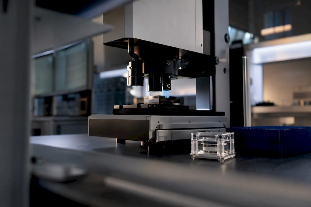
AMPK activators split into two mechanistic classes: direct ligands like A-769662 and MK-8722 that bind the ADaM site and switch on the kinase independent of cellular energy state, and indirect agents like metformin and berberine that work by raising the AMP:ATP ratio through mild energy stress. Both classes show real therapeutic promise in metabolic disease, but isoform and tissue specificity determine whether that promise turns into benefit or into off-target harm, since systemic pan-activators such as MK-8722 have produced cardiac hypertrophy in preclinical models.
TL;DR:
- Systemic pan-activators like MK-8722 can cause cardiac hypertrophy due to their activation of all AMPK isoforms in cardiac tissue, posing safety concerns.
- Direct activators such as A-769662 and compound 991 engage the ADaM site in vitro, but their tissue specificity is limited by isoform and subunit composition, influencing their research and therapeutic relevance.
- Natural compounds like berberine and resveratrol activate AMPK indirectly through mitochondrial stress, but their effectiveness at achievable doses in humans remains uncertain.
- Validating AMPK activation mechanisms requires cell-free assays, measuring nucleotide ratios, and confirming dependence on the ADaM site, especially when evaluating new compounds.
- Designing tissue- or isoform-selective agents, combined with early safety testing, is a safer, more promising approach than developing broad-spectrum activators with systemic effects.
Synthrolab
Support Rigorous AMPK Research
Synthro Lab provides research-grade peptides and laboratory compounds for investigating cellular signalling, metabolism, and longevity-related mechanisms.
Explore research compounds
Table of Contents
What Are AMPK Activators and How Do They Work?
AMP-activated protein kinase is a heterotrimeric enzyme built from a catalytic α-subunit, a scaffolding β-subunit, and a regulatory γ-subunit. Each piece has a job. The α-subunit carries the kinase domain (KD) and the critical Thr172 residue. The β-subunit contains a carbohydrate-binding module (CBM) and, right next to it, a shallow pocket formed at the interface of the KD and CBM. That pocket is the ADaM site, short for allosteric drug and metabolite site, and it is the single most important structural feature in this entire field.
The γ-subunit binds AMP, ADP, and ATP competitively. When cellular energy drops, the (AMP+ADP):ATP ratio climbs, and AMP or ADP displacement of ATP on the γ-subunit triggers two things: it makes Thr172 phosphorylation by upstream kinases more likely, and it protects that phosphate group from removal by protein phosphatases. Thr172 sits on the activation loop of the kinase domain, and its phosphorylation status is the master switch. Two upstream kinases do the phosphorylating: LKB1, which handles most of the constitutive, energy-sensing activation, and CaMKKβ, which responds to calcium spikes independent of energy charge. This is why calcium-mobilizing natural compounds can activate AMPK even when ATP levels look normal.
Direct activators skip the nucleotide-sensing step entirely. Compounds like A-769662 wedge into the ADaM site and physically stabilize the interaction between the kinase domain and the CBM, locking the activation loop into a conformation that resists dephosphorylation. Crystal structures of AMPK bound to A-769662 and the related compound 991 show exactly this: the ligand acts like a molecular clamp holding two domains together that would otherwise drift apart, according to the mechanistic review published in Frontiers in molecular biosciences. That structural insight explains why direct activators can turn on AMPK in cell-free systems with no ATP depletion at all.
A few structural facts matter for anyone designing experiments around this pocket:
- The ADaM site only exists in a fully assembled heterotrimer, so isolated kinase domain constructs will not respond to A-769662 or 991.
- Ligand occupancy at the ADaM site increases Thr172 phosphorylation stoichiometry by protecting it from phosphatases, not primarily by recruiting more upstream kinase.
- β1 and β2 isoforms differ in ADaM pocket sequence, which is why several ADaM ligands show strong β1 preference and weak or absent activity at β2-containing complexes.
- Nucleotide-based activation (AMP, ADP) and ADaM-site activation are additive rather than mutually exclusive, meaning a cell under real energy stress plus a direct activator can show synergistic Thr172 phosphorylation.
Get the subunit composition of your target complex wrong, and a compound that looks inactive may simply be hitting the wrong isoform pairing.
Direct vs. Indirect Activators: Telling Them Apart in the Lab
Classifying a compound as direct or indirect is not a matter of reading the label on a vial. It comes down to whether the molecule engages the ADaM site on purified, reconstituted AMPK in the total absence of nucleotide flux, or whether it needs an intact cell with a functioning mitochondrion and energy-sensing machinery to produce any effect at all.
- Run a cell-free activation assay first. Purified α/β/γ heterotrimer plus a compound, with ATP substrate and no cellular context, will activate only in response to true direct binders. If a candidate shows zero activity here but strong activity in intact cells, you are looking at an indirect mechanism.
- Measure the AMP:ADP:ATP ratio in treated cells. Indirect activators like metformin, berberine, and AICAR shift this ratio measurably, usually within thirty to sixty minutes, because they interfere with oxidative phosphorylation or mimic AMP directly. Direct ADaM binders leave the nucleotide ratio essentially untouched even while Thr172 phosphorylation rises.
- Track p-Thr172 and p-ACC together. Acetyl-CoA carboxylase phosphorylation at Ser79 is the standard downstream readout, and it should track with p-Thr172 regardless of mechanism. A mismatch, where ACC phosphorylation lags far behind reported Thr172 signal, often flags an antibody artifact or an off-target kinase rather than genuine AMPK engagement.
- Test dependence on mitochondrial function. Block oxidative phosphorylation independently (oligomycin, rotenone) and see whether the candidate compound’s activity is additive or redundant with that block. Indirect activators that work through complex I inhibition, like metformin, will show redundancy; direct binders will not.
- Confirm ADaM-site dependence with mutant constructs. A β1 S108A mutation abolishes A-769662 and 991 activity almost completely, which is the gold-standard genetic test for ADaM-site engagement.
Mapped against these tests, the canonical direct activators are A-769662, compound 991, and MK-8722, all of which engage the ADaM pocket in cell-free assays. The canonical indirect activators are metformin (mild complex I inhibition), berberine (similar mitochondrial mechanism plus additional pathways), and AICAR, which is converted intracellularly to ZMP, an AMP mimetic that binds the γ-subunit’s nucleotide sites directly rather than the ADaM pocket on the β-subunit. AICAR’s dependence on cellular adenosine kinase for conversion also means its potency varies significantly by cell type, something worth checking before assuming a null result reflects target biology rather than poor prodrug conversion.
Small-Molecule AMPK Activators: Potency, Selectivity, and Safety Signals
A-769662 was the first widely used ADaM-site ligand and remains a benchmark tool compound, but it carries real limitations: modest cell permeability, off-target inhibition of certain ion channels at higher concentrations, and strict β1-selectivity that makes it nearly useless for probing β2-dominant tissue like skeletal muscle. Compound 991 improved on potency and binds the same pocket with a similar β1 bias, giving researchers a slightly cleaner tool for liver and other β1-rich tissues work.
MK-8722 changed the conversation. Unlike its ADaM predecessors, it activates all AMPK complexes regardless of β-isoform, making it a true systemic pan-activator. In preclinical studies, MK-8722 produced sustained glucose lowering after a single dose, an efficacy signal strong enough to draw serious pharmaceutical interest. But the same paper that established its potency also reported cardiac hypertrophy in treated research subjects, a finding documented in the discovery paper published through PMC.
Pro Tip: If you’re evaluating a pan-activator for a metabolic disease model, build cardiac histology and echocardiography into the protocol from day one rather than adding it retroactively after you see a glucose effect worth chasing.
That cardiac finding is arguably the single most important data point in this field for anyone doing translational work, because it demonstrates that broad, isoform-blind activation of a kinase this central to cellular energy metabolism does not come free. AMPK activation in cardiac tissue drives glycogen accumulation, and sustained pan-activation appears to push that process past a tolerable threshold into pathological hypertrophy.
A short comparison of the core direct-activating small molecules:
- A-769662: β1-selective, requires Ser108 on β1 for activity, useful as a mechanistic tool compound but poor translational candidate due to permeability and selectivity limits.
- Compound 991: Similar β1 bias to A-769662 with improved potency, still primarily a research tool rather than a clinical candidate.
- MK-8722: Pan-isoform activator, strong glucose-lowering efficacy, but flagged for cardiac hypertrophy risk with chronic systemic exposure.
- AICAR: An AMP mimetic rather than an ADaM binder, useful experimentally but requires intracellular conversion to ZMP and shows highly variable potency across cell types.
The lesson across all four compounds is consistent: potency and selectivity move independently, and a molecule that activates AMPK powerfully everywhere in the body is not automatically the better research or therapeutic candidate compared to one that activates it powerfully in one tissue.
Natural and Nutraceutical AMPK Activators: What the Evidence Actually Supports
Several plant-derived and dietary compounds activate AMPK, but almost none of them do it through the ADaM site the way A-769662 does. Most work through a mix of mild mitochondrial stress, calcium signaling, or, in one notable case, a completely different allosteric surface.
Resveratrol activates AMPK indirectly, largely through mild inhibition of ATP synthase that raises the cellular AMP:ATP ratio, and shows cardioprotective and metabolic benefits in animal models. The catch, well documented in a review of natural AMPK activators in cardiovascular disease, is that the concentrations required for reliable AMPK activation in vitro frequently exceed what oral dosing achieves in human plasma, given resveratrol’s poor bioavailability and rapid glucuronidation.
Berberine works through a comparable mitochondrial mechanism and has the strongest human epidemiological and clinical trial backing among the botanicals, largely because it has been studied as a glucose-lowering agent independent of its AMPK activity, giving researchers real pharmacokinetic data to work from.
Salicylate, the active metabolite of aspirin, is the outlier on this list because it does not work like the other natural compounds at all. Salicylate binds directly to the same ADaM pocket that A-769662 occupies and produces a modest, roughly 10 to 15 percent increase in AMPK activity at clinically relevant concentrations, acting complementarily to AMP rather than replacing it, according to findings summarized in work on AMPK activation and healthspan. That effect size is real but small, and it is a useful reminder that even direct ADaM engagement does not guarantee dramatic activation.
Vinegar and acetate raise AMPK activity through a slightly different route: acetate gets converted to acetyl-CoA and ultimately shifts cellular AMP:ATP balance, though the effect is generally weaker and more transient than what mitochondrial inhibitors produce.
Quercetin shows AMPK-activating effects in cell culture, but the evidence for meaningful in vivo activation at achievable dietary or supplemental intake remains thin compared to berberine or resveratrol.
A few practical notes for using these agents as experimental probes:
- Treat resveratrol, berberine, and quercetin as tools for hypothesis generation in vitro, not as calibrated activators with known human-relevant potency.
- Confirm any observed AMPK activation is not simply general mitochondrial toxicity by pairing treatment with an OCR (oxygen consumption rate) assay.
- Recognize that computational docking studies, including recent work screening fenugreek and oyster mushroom compounds for ADaM-site binding, can generate promising candidates, but predicted binding affinity means little without wet-lab validation.
- Report the exact concentration used relative to published human plasma achievable levels whenever claiming physiological relevance for a natural compound.
Where AMPK Activation Helps, and Where It Can Hurt
Metabolic disease is where AMPK activators have the deepest evidence base. Metformin’s decades of clinical use and berberine’s growing trial data both point toward genuine glucose-lowering and lipid benefits tied at least partly to AMPK-dependent mechanisms, though metformin also acts through AMPK-independent pathways in the liver that complicate clean mechanistic attribution.
Cardiovascular disease presents a genuinely two-sided picture. Acute, transient AMPK activation during exercise or intermittent metabolic stress supports healthy cardiac metabolism and is broadly protective. Chronic, high-level pan-activation is a different story. The MK-8722 cardiac hypertrophy finding is the clearest warning sign in the literature: sustained activation across all AMPK isoforms in cardiac tissue drove glycogen accumulation severe enough to produce structural hypertrophy in preclinical models. That single result should temper enthusiasm for any systemic, isoform-blind activator intended for long-term use.
Cancer research tells a genuinely complicated story rather than a simple good-or-bad one. AMPK can suppress tumor growth by restricting anabolic processes tumor cells need to proliferate, but in some contexts, tumor cells co-opt AMPK activity to survive nutrient stress within the tumor microenvironment. The ACS medicinal chemistry review covering small-molecule AMPK modulators in oncology stresses that preclinical models frequently fail to replicate the nutrient-deprived, hypoxic conditions of real tumors, which can make an activator look uniformly beneficial in a well-oxygenated culture dish while behaving very differently in vivo.
Aging and healthspan research treats AMPK as one node in a broader energy-sensing network alongside mTOR and sirtuins, with transient activation patterns (fasting, exercise, caloric restriction) associated with favorable metabolic outcomes across model organisms. This is the softest evidence category on the list. Extrapolating from lifespan studies in simple organisms to pharmacological human intervention remains speculative.
For any translational program working with systemic activators, a minimal safety endpoint panel should include:
- Cardiac imaging (echocardiography) and histology to catch hypertrophy or glycogen accumulation early.
- Direct cardiac glycogen quantification rather than relying on imaging alone.
- HbA1c and fasting glucose to track the metabolic effect the compound is presumably being developed for.
- A full lipid panel, since several indirect activators alter lipid metabolism independent of glucose effects.
AMPK exists as multiple complexes built from two α-isoforms (α1, α2), two β-isoforms (β1, β2), and three γ-isoforms (γ1, γ2, γ3), and these combinations are not evenly distributed across the body. α2-containing complexes dominate in skeletal muscle and are the primary drivers of exercise-induced metabolic adaptation. α1 is more broadly expressed and dominant in tissues like the liver and immune cells. β2 is the primary isoform in skeletal muscle and heart, while β1 dominates in liver, which directly explains why β1-selective ADaM binders like A-769662 barely touch cardiac or muscle AMPK activity at all.
That tissue distribution is precisely why MK-8722’s pan-isoform activity, rather than being a strength, turned into its central liability. A β1-selective compound would likely never have produced the cardiac signal MK-8722 did, because β2 dominates heart tissue and a β1-only ligand would largely leave it alone.
Practical strategies researchers are pursuing to improve the therapeutic window:
- Medicinal chemistry optimization targeting sequence differences in the ADaM pocket between β1 and β2 to achieve tissue-restricted activation.
- Tissue-targeted delivery systems (conjugation, nanoparticle encapsulation, or tissue-specific prodrugs) that concentrate exposure in liver or muscle while sparing cardiac tissue.
- Combination dosing at sub-maximal levels of a pan-activator alongside a tissue-restricting delivery vehicle, rather than relying on isoform selectivity alone.
Before drawing conclusions from any AMPK activation study, confirm exactly which isoform combinations the model system expresses. A hepatocyte line dominated by β1 will give a misleadingly clean result with A-769662 that would never replicate in cardiac or skeletal muscle tissue, and reporting that result without the isoform context invites exactly the kind of overgeneralization the field has spent the last decade trying to correct, a point the ACS review makes explicit when discussing why isoform-specific modulators are now the preferred research direction.
Building a Reproducible AMPK Activation Experiment
Most failed replications in this field trace back to three overlooked variables: cell line LKB1 status, baseline energy state, and oxygenation. Skip any one of them and a promising result can vanish the moment another lab tries to reproduce it.
- Verify LKB1 expression before starting. Several commonly used cancer cell lines carry LKB1 mutations, and since LKB1 handles the majority of canonical Thr172 phosphorylation, a compound tested in an LKB1-null line may show a falsely blunted response that has nothing to do with its actual mechanism.
- Standardize baseline energy state and oxygenation. High-glucose, high-oxygen culture conditions common in standard cell culture protocols do not reflect physiological energy stress, and this mismatch is one of the more consistent sources of failed in vitro to in vivo translation.
- Include primary cells or in vivo models wherever the research question demands tissue relevance. Immortalized lines are convenient but frequently diverge from primary tissue in both isoform expression and baseline metabolic state.
- Run the full assay panel, not a single readout. Kinase activity assays, p-Thr172 western blotting, p-ACC as a downstream functional check, oxygen consumption rate via Seahorse or equivalent, and direct AMP:ADP:ATP quantification together give a far more trustworthy picture than any single measurement alone.
- Screen for off-target mitochondrial toxicity. Many indirect activators work by perturbing oxidative phosphorylation, and a compound can produce AMPK activation as a side effect of general mitochondrial poisoning rather than a targeted mechanism worth pursuing further.
Pro Tip: Run a concentration-response curve alongside a fixed, published reference concentration for your comparator compound. Reporting your novel candidate’s EC50 without that anchor makes cross-study comparison nearly impossible for anyone reading your data later.
Concentration mismatches deserve special attention. A compound that activates AMPK cleanly at 50 micromolar in a dish frequently has no realistic path to that exposure level in a living organism, and treating an in vitro concentration as if it predicts in vivo relevance is one of the more common overreach errors in published AMPK work. Synthro Lab’s guides on interpreting basic lab research findings and metabolic health markers walk through how to set these assay panels up and read the results without overinterpreting a single positive signal.
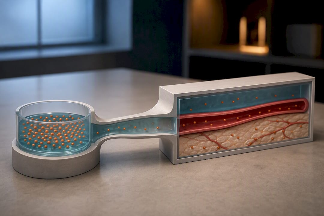
The field has spent the last several years correcting an early enthusiasm for potent, broad-spectrum AMPK activators, and I think that correction is overdue rather than excessive. MK-8722’s cardiac finding was not a fluke. It was a predictable consequence of activating a kinase that regulates cardiac glycogen everywhere at once, with no tissue brake on the effect.
The more promising direction, and the one worth the field’s attention over the next several years, is isoform-selective and tissue-targeted delivery, not incrementally more potent pan-activators. Short-term experimental programs should prioritize combination probes that pair modest indirect activation with tissue-restricted direct ligands, alongside cardiac and hepatic safety screens run in parallel from the earliest preclinical stage rather than bolted on after efficacy looks promising. Synthro Lab’s resources on metabolic health compound classes go deeper into how these categories map onto research design.
— Mitch
Sourcing Research-Grade Compounds for AMPK Studies
Running the kind of AMPK activation study outlined here means sourcing compounds you can trust down to the batch, since a contaminated or mislabeled reagent will cost you weeks chasing an artifact instead of a real signal. Research-grade peptides and laboratory compounds with independent batch testing and certificates of analysis are essential when trying to distinguish a true ADaM-site effect from off-target mitochondrial noise.
Synthrolab’s catalog covers the metabolic and cellular signaling categories relevant to AMPK-adjacent research, including compounds researchers pair with metabolic studies when investigating downstream signaling and recovery pathways. If your protocol involves GLP-related metabolic compounds alongside AMPK work, the GLP 1 (Sema) product page has full specifications and COA access. For a broader look at how these compound categories fit together in a metabolic research program, start with the metabolic health compounds overview and check current lot documentation before placing your next order.
This article is general information, not a substitute for advice from a qualified doctor. Consult a qualified healthcare professional about your own circumstances before acting on anything here.
Sources
FAQ
What Are the Best AMPK Activators for Research Use?
There is no single best activator. It depends on the question: A-769662 and compound 991 are the standard tools for β1-selective ADaM-site engagement, MK-8722 is the reference pan-activator for systemic efficacy studies, and metformin, berberine, or AICAR are the go-to indirect agents for energy-stress models.
Do AMPK Activators Actually Work?
Direct activators like A-769662 and MK-8722 reliably activate AMPK in cell-free and cellular assays, and indirect agents like metformin have decades of clinical data behind their metabolic effects, but “working” pharmacologically is not the same as being risk-free, since MK-8722’s preclinical cardiac hypertrophy finding shows potent activation can carry real tissue-specific costs.
How Can You Naturally Activate AMPK?
Exercise and intermittent fasting are the best-supported natural triggers, since both raise the cellular AMP:ADP:ATP ratio through genuine energy demand. Dietary compounds like berberine and resveratrol also activate AMPK in preclinical models, though achievable human plasma concentrations often fall below what full in vitro activation requires.
What Foods Contain AMPK Activators?
No food delivers a pure ADaM-site ligand, but several contain compounds with AMPK-activating properties in preclinical studies: berberine-containing plants, resveratrol-rich sources like red grape skin, and acetate from vinegar all show measurable, if modest, activation effects.
What Is the Difference Between AICAR and Direct ADaM-Site Activators?
AICAR converts intracellularly to ZMP, an AMP mimetic that binds the γ-subunit’s nucleotide-sensing sites, while direct activators like A-769662 and MK-8722 bind the separate ADaM pocket on the β-subunit and activate the kinase independent of nucleotide levels entirely.
Recommended
 21+ verification