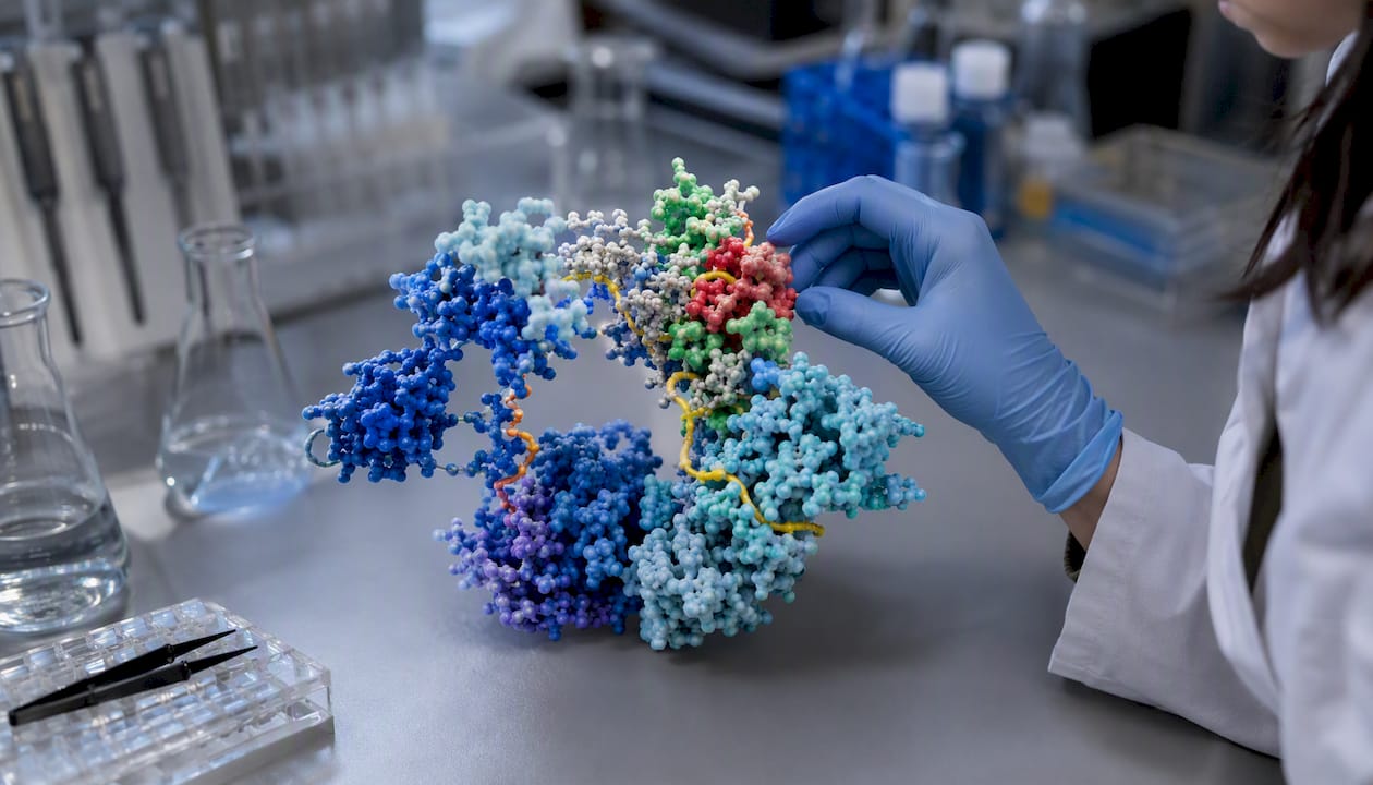
Cellular signalling is the process by which cells detect, interpret, and respond to chemical or physical information from their environment, neighboring cells, or distant tissues. Every signalling event involves three core components: a signalling molecule (ligand), a receptor protein that recognizes it, and a signal transduction cascade that converts the binding event into a cellular action. As Wikipedia’s cell signaling entry frames it, the process spans from the first messenger all the way through the transduction machinery to the final biological response.
These molecular conversations control nearly everything a cell does: whether it divides, differentiates into a specialized type, ramps up metabolism, or triggers its own death. When the system breaks down, the consequences are serious. Dysregulated signalling underlies cancer, type 2 diabetes, autoimmune disorders, and neurodegenerative disease. One figure captures just how sensitive this system is:
Statistic: Signalling molecules often act at concentrations at or below 10⁻⁸ M, and their receptors bind them with affinities of Ka ≥ 10⁸ L/mol. A vanishingly small amount of ligand can trigger a large, amplified response inside the cell.
Authoritative sources for this article include the NCBI Bookshelf editions of Molecular Biology of the Cell, Nature Scitable’s cell signaling topic page, and StatPearls.
Key Takeaways
Cellular signalling is the molecular language cells use to coordinate every biological process, from metabolism to development to immune defense, and its dysregulation drives most major diseases.
| Point |
Details |
| Core definition |
Cellular signalling involves a ligand, a receptor, and a transduction cascade converting an external cue into a cellular response. |
| Three-stage framework |
Reception, transduction, and response are the universal stages of every signalling event, regardless of pathway. |
| Receptor class determines mechanism |
GPCRs use second messengers; RTKs use phosphorylation cascades; intracellular receptors regulate transcription directly. |
| Amplification and cross-talk |
A single receptor activation can phosphorylate thousands of targets; pathways intersect, so blocking one node often affects others. |
| Clinical relevance |
Dysregulated signalling underlies cancer, type 2 diabetes, autoimmune disorders, and neurodegenerative disease. |
Table of Contents
What is cellular signalling, and how does it work step by step?
Every signalling event follows the same three-stage sequence: reception → transduction → response. Understanding each stage is the fastest way to read any pathway diagram or pharmacology paper with confidence.
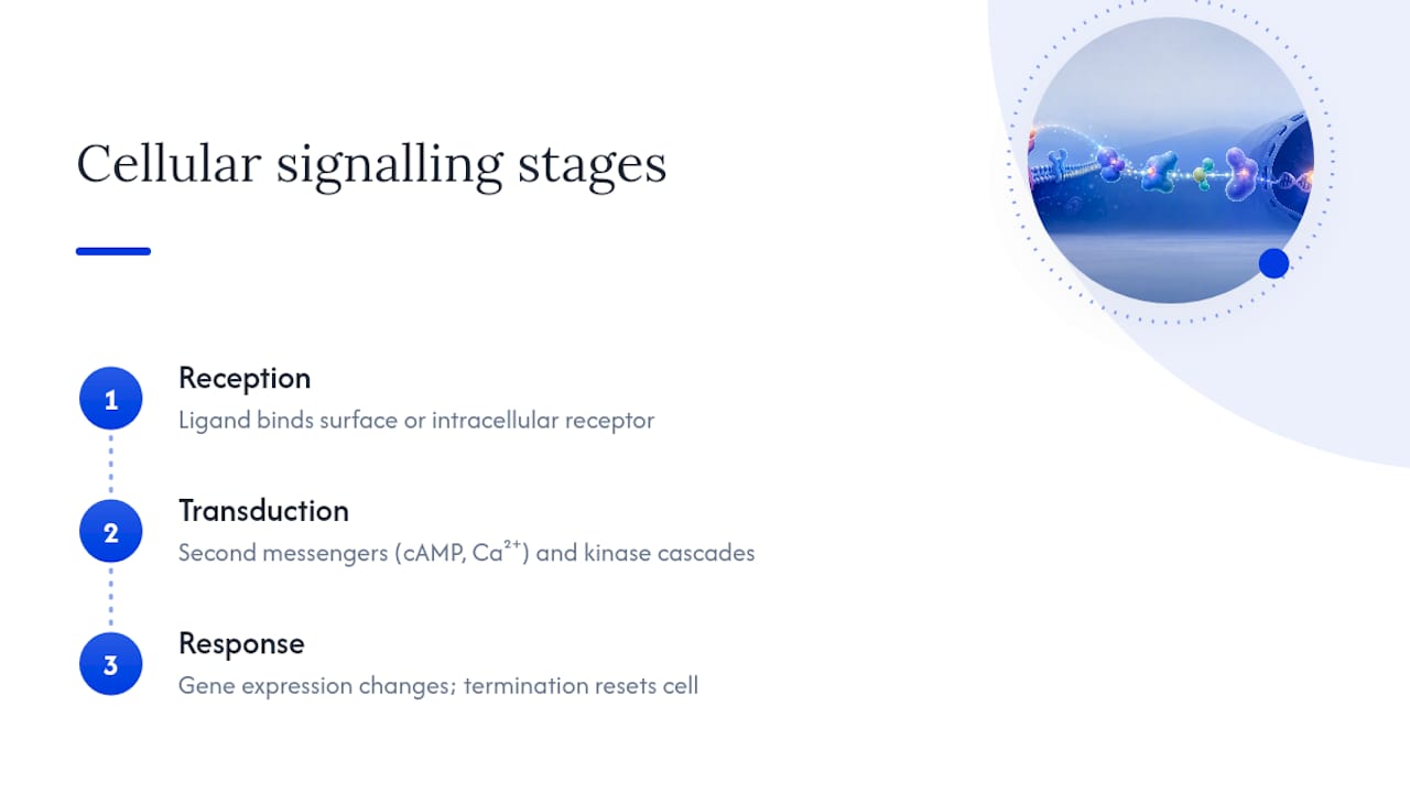
Reception is the moment a ligand binds its receptor. The receptor can sit on the cell surface (for water-soluble messengers like peptide hormones) or inside the cell (for hydrophobic messengers like steroid hormones that diffuse through the plasma membrane). Binding is highly specific: a receptor recognizes its ligand the way a lock recognizes a key, and that specificity is what keeps signals from crossing wires.
Transduction is where the chemistry gets interesting. The receptor’s conformational change launches a chain of intracellular events, often involving second messengers such as cyclic AMP (cAMP) or calcium ions (Ca²⁺), and kinase cascades that phosphorylate downstream proteins. Each step in the cascade can amplify the signal, so a single receptor activation can ultimately phosphorylate thousands of target proteins. Cells can also communicate by direct contact through gap junctions, which permit direct cytoplasmic exchange between connected cells, bypassing the need for a secreted messenger entirely.
Response is the outcome: a change in gene expression, a metabolic shift, cytoskeletal reorganization, or the activation of a secretory program. The response is not permanent. Signal termination mechanisms, including receptor internalization, phosphatase activity that reverses phosphorylation, and enzymatic degradation of second messengers, reset the system so the cell can respond again. Without termination, a cell would be stuck in a permanently activated state, which is exactly what happens in some cancers.
Key terms at a glance
| Term |
Plain-language definition |
| Ligand |
The signalling molecule that binds a receptor to start a cascade |
| Receptor |
A protein that recognizes a specific ligand and changes shape upon binding |
| Second messenger |
A small intracellular molecule (e.g., cAMP, Ca²⁺) that relays the signal inside the cell |
| Kinase |
An enzyme that adds a phosphate group to a target protein, activating or inactivating it |
| Signal transduction |
The full chain of molecular events between ligand binding and cellular response |
| Desensitization |
Reduced receptor responsiveness after prolonged or repeated stimulation |
Diagram suggestion: a labeled three-panel figure showing (1) ligand binding at the cell surface, (2) second-messenger generation and kinase cascade, and (3) nuclear transcription factor activation would clarify the spatial flow for visual learners.
How signals reach their targets: the five modes of cell communication
Signalling molecules are classified by how far they travel and how they are delivered. The target cell must express a complementary receptor, or the signal simply goes unheard.
Autocrine signalling: A cell secretes a messenger that binds receptors on its own surface. Certain immune cells, for example, release interleukins that stimulate their own proliferation during an immune response. In cancer, autocrine loops can become self-sustaining growth signals.
Juxtacrine signalling: Requires direct cell-to-cell contact. The ligand stays membrane-bound on the sending cell and engages a receptor on an immediately adjacent cell. Notch-Delta signalling during neuronal development is a classic example, where the outcome depends entirely on which cell happens to be touching which.
Paracrine signalling: The messenger diffuses a short distance through the extracellular space to act on nearby cells. Growth factors released at a wound site act parabolically, stimulating local fibroblasts and endothelial cells without flooding the bloodstream.
Endocrine signalling: Hormones travel through the bloodstream to reach distant target tissues. Insulin, secreted by pancreatic beta cells, acts on muscle, liver, and fat tissue across the entire body. The trade-off for that reach is speed: endocrine signals are slower than paracrine or synaptic ones.
Synaptic (neuronal) signalling: A hybrid of electrical and chemical mechanisms. An action potential travels down a neuron’s axon, triggers neurotransmitter release into the synaptic cleft, and the neurotransmitter binds postsynaptic receptors within milliseconds. The speed and precision here are unmatched by any other signalling mode.
| Dimension |
Autocrine |
Juxtacrine |
Paracrine |
Endocrine |
Synaptic |
| Range |
Same cell |
Adjacent cell |
Local tissue |
Whole organism |
Synapse (nanoscale) |
| Speed |
Fast |
Fast |
Moderate |
Slow |
Extremely fast |
| Delivery route |
Secreted, self-binding |
Membrane-bound ligand |
Diffusion |
Bloodstream |
Neurotransmitter release |
| Example |
IL-2 in T cells |
Notch-Delta in neurons |
Wound-site growth factors |
Insulin |
Acetylcholine at NMJ |
| Typical receptor |
Surface or intracellular |
Surface (Notch family) |
RTKs, GPCRs |
GPCRs, nuclear |
Ionotropic, metabotropic |
The four major receptor families and how they transduce signals
After ligand binding, three main cell-surface receptor families handle most of the transduction work: GPCRs, ion-channel receptors, and enzyme-linked receptors. A fourth class, intracellular receptors, handles hydrophobic messengers that cross the membrane on their own.
G-protein-coupled receptors (GPCRs) are the largest receptor superfamily in the human genome, with over 800 members. When a ligand binds, the receptor activates a heterotrimeric G-protein, which exchanges GDP for GTP and then dissociates to modulate downstream enzymes (adenylyl cyclase, phospholipase C) or ion channels. The result is a burst of second messengers. Because GPCRs sit upstream of so many pathways, roughly 34% of all FDA-approved drugs target them, from beta-blockers to antihistamines.
Ion-channel-linked receptors (ionotropic receptors) open or close an ion channel directly upon ligand binding. The response is measured in milliseconds, making these the go-to receptors for fast synaptic transmission. Nicotinic acetylcholine receptors at the neuromuscular junction are the textbook example: acetylcholine binding opens a cation channel, sodium floods in, and the muscle depolarizes.
Enzyme-linked receptors, particularly receptor tyrosine kinases (RTKs), dimerize upon ligand binding and phosphorylate each other’s intracellular domains. Those phosphotyrosine sites then recruit adaptor proteins and activate cascades such as the MAPK and PI3K-Akt pathways, which drive cell growth and survival. Epidermal growth factor receptor (EGFR) is an RTK that is mutated or overexpressed in several cancers, making it a major drug target.
Intracellular (nuclear) receptors bind hydrophobic ligands, including steroid hormones, thyroid hormone, and retinoic acid, that diffuse across the plasma membrane. Once bound, the receptor-ligand complex typically translocates to the nucleus and acts directly as a transcription factor. The response is slower than surface-receptor signalling but can be long-lasting because it rewires gene expression programs.
Detecting receptor activation in the lab
- Ligand-binding assays: Radiolabeled or fluorescent ligands quantify receptor occupancy and affinity.
- Phosphorylation Western blots: Antibodies against phosphorylated residues (e.g., pTyr, pSer) confirm RTK or kinase activation.
- cAMP reporter assays: HTRF or BRET-based kits measure GPCR-driven cAMP changes in real time.
- Calcium imaging: Fluorescent dyes (Fura-2, Fluo-4) report intracellular Ca²⁺ flux with subcellular resolution.
- Co-immunoprecipitation: Pulls down receptor-adaptor complexes to confirm protein-protein interactions downstream of activation.
Inside the cell: second messengers, kinase cascades, and cross-talk
A single activated receptor can generate thousands of second-messenger molecules, and each of those can activate multiple kinases. That amplification logic is why signalling cascades of protein kinases and second messengers allow one receptor activation to trigger large cellular responses.
cAMP is produced by adenylyl cyclase after GPCR activation. It activates protein kinase A (PKA), which phosphorylates transcription factors like CREB, ultimately changing which genes are expressed. Ca²⁺ released from the endoplasmic reticulum activates calmodulin-dependent kinases and triggers processes ranging from muscle contraction to neurotransmitter release. Diacylglycerol (DAG) activates protein kinase C (PKC), which feeds into the MAPK cascade and regulates proliferation.
The MAPK cascade is a good example of amplification in action: Ras → Raf → MEK → ERK, with each step phosphorylating and activating the next. ERK then enters the nucleus and phosphorylates transcription factors that drive cell cycle progression.
Cross-talk between pathways is the rule, not the exception. PKA can phosphorylate components of the MAPK pathway; Ca²⁺ can modulate adenylyl cyclase activity. Downstream target proteins are common currency across many pathways, so different upstream signals can converge on the same effectors. For researchers, this means blocking one node often reroutes flux through a parallel pathway, producing unexpected phenotypes.
Example pathway: GPCR → transcriptional effect
| Step |
Molecule/Event |
| 1. Ligand binds GPCR |
Receptor changes conformation |
| 2. G-protein activation |
GDP → GTP exchange; Gα dissociates |
| 3. Adenylyl cyclase activated |
ATP → cAMP |
| 4. PKA activated |
cAMP binds regulatory subunits |
| 5. CREB phosphorylated |
PKA enters nucleus |
| 6. Gene transcription |
CREB binds CRE promoter elements |
Statistic: Because receptors bind ligands at concentrations at or below 10⁻⁸ M, assay sensitivity must match the pathway’s amplification profile. A fast ionic event (milliseconds) and a slow transcriptional response (hours) require completely different experimental designs.
Real-world examples and what goes wrong when signalling fails
Mechanisms become memorable when you can attach them to something familiar.
1. Insulin and metabolic regulation. Insulin binds the insulin receptor (an RTK) on muscle and fat cells, triggering glucose transporter (GLUT4) translocation to the cell surface. In type 2 diabetes, downstream components of this pathway become desensitized, so cells stop responding even when insulin is present. For more on how this connects to energy metabolism, the metabolic signalling context is worth exploring.
2. Neurotransmitters and synaptic signalling. Glutamate activates both ionotropic receptors (fast, direct ion flux) and metabotropic receptors (slower, GPCR-mediated) in the brain. Imbalances in glutamate signalling are implicated in epilepsy, schizophrenia, and excitotoxic neuronal death after stroke.
3. Growth factors and cell proliferation. EGF binding to EGFR activates the MAPK cascade and drives cell division. EGFR mutations that lock the receptor in a constitutively active state are found in a significant proportion of non-small-cell lung cancers, making EGFR a validated drug target for tyrosine kinase inhibitors.
4. Autocrine loops in cancer. Some tumor cells secrete their own growth factors and express the matching receptors, creating a self-sustaining proliferation signal that bypasses normal tissue controls.
5. Nitric oxide as a paracrine gaseous messenger. Endothelial cells produce nitric oxide (NO), which diffuses into adjacent smooth muscle cells and activates guanylyl cyclase, raising cGMP and causing relaxation. Nitroglycerin works by releasing NO and dilating coronary vessels.
6. Developmental signalling and differentiation. Cell signalling coordinates the gene-expression programs that convert populations of identical progenitor cells into organized tissues and organs. Wnt, Hedgehog, and Notch pathways are the primary architects of body-plan patterning. Mutations in any of them can cause developmental defects or pediatric cancers. Senescent cells illustrate the flip side: their secretory signalling (SASP) actively disrupts tissue homeostasis in aging.
Signalling errors are not a modern phenomenon. Evolutionary evidence shows that unicellular organisms used intercellular signalling before multicellularity evolved; present-day yeasts secrete mating factors that are structurally analogous to mammalian peptide hormones. The core logic of ligand-receptor-cascade is ancient.
Diseases tied to signalling dysregulation:
- Cancer (constitutively active RTKs, loss of tumor suppressor signalling)
- Type 2 diabetes (insulin receptor pathway desensitization)
- Autoimmune disorders (aberrant cytokine and T-cell receptor signalling)
- Cardiovascular disease (impaired NO-cGMP signalling in endothelium)
- Neurodegenerative disease (disrupted neurotrophin and glutamate signalling)
How scientists study cellular signalling
The core goal of most signalling experiments is to link receptor activation to a measurable intracellular change, then establish causality by perturbing one node and observing what shifts. Here are the main tools researchers reach for:
Ligand-binding assays quantify receptor affinity and occupancy using radiolabeled or fluorescent ligands. They answer the first question: does the compound actually bind?
Reporter assays (luciferase, SEAP, or BRET-based cAMP kits) measure pathway output in living cells without lysing them, making them ideal for kinetic studies.
Western blotting for phosphoproteins is the workhorse for confirming kinase cascade activation. Phospho-specific antibodies against pERK, pAkt, or pSTAT proteins give a snapshot of pathway state at a defined time point.
Fluorescence calcium imaging with dyes like Fura-2 or genetically encoded indicators (GCaMP) captures Ca²⁺ dynamics in real time, essential for studying fast signalling in neurons or cardiomyocytes.
FRET and BRET biosensors report protein-protein interactions or conformational changes in living cells with spatial and temporal precision that lysate-based assays cannot match.
Mass spectrometry-based phosphoproteomics maps thousands of phosphorylation sites simultaneously, giving a system-wide view of which kinases are active under a given condition.
RNA-seq captures downstream transcriptional responses, connecting receptor activation to gene-expression changes hours or days later.
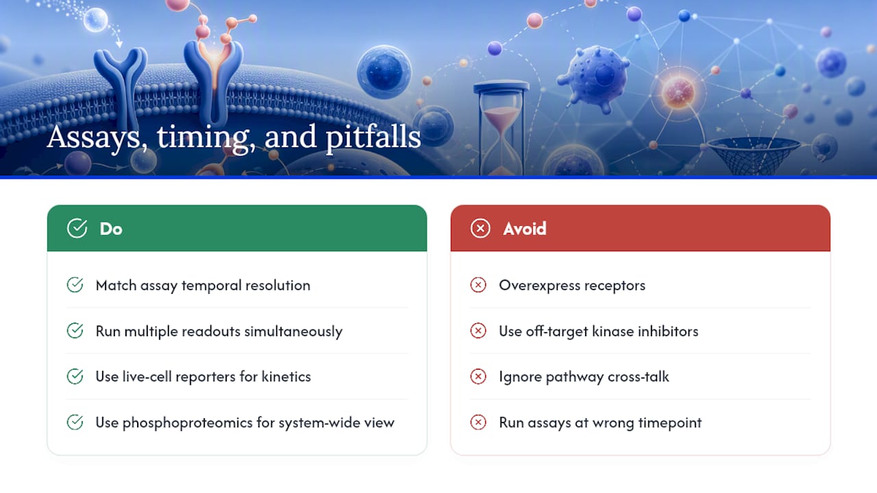
Pro Tip: Match your assay’s temporal resolution to the pathway you are studying. A fast ionotropic event resolves in milliseconds; a transcriptional response peaks in hours. Running a Western blot at the wrong time point is one of the most common reasons a real signalling event goes undetected. For guidance on reading and interpreting these experimental outputs, Synthrolab’s researcher guide covers common pitfalls in detail.
Common pitfalls include overexpression artifacts (transfected receptor levels far exceed physiological levels), off-target pharmacology (many kinase inhibitors hit multiple kinases at the concentrations used), and pathway cross-talk producing unexpected phenotypes when one node is blocked. Running multiple readouts simultaneously, rather than relying on a single assay, is the most reliable way to build a coherent picture.
Why signalling knowledge shapes every research decision
Understanding cellular signalling is not background reading you do once and forget. It is the interpretive framework that determines which experiment to run next, which control to include, and which result to trust.
The modularity of signalling pathways means that a peptide or compound acting on one receptor can ripple through multiple downstream networks. Synthrolab supplies research-grade peptides and laboratory compounds, each independently batch-tested with certificates of analysis, specifically for researchers investigating these mechanisms: cellular signalling, metabolism, recovery pathways, mitochondrial function, and longevity-related biology. For researchers exploring how specific peptides interact with signalling networks, the stem cell signalling peptides overview and the PGC-1 alpha pathway explainer are practical starting points.
The deeper you understand the receptor-to-response logic, the better your experimental design will be, and the less likely you are to mistake a cross-talk artifact for a primary effect.
Exploring research-grade compounds for signalling studies
Researchers who want to probe specific signalling pathways need compounds with verified purity. Synthrolab’s catalog includes peptides relevant to cellular regeneration, anabolic signalling, and longevity mechanisms, all sold for research use only. The GHK-Cu + BPC-157 + TB-500 bundle is one example of compounds studied for their interactions with repair and regeneration signalling. For researchers newer to peptide work, Peptides for Beginners explains quality metrics and reconstitution basics before you run your first assay.
Sources
The sources below are the primary references used in this article, selected for authority and accessibility.
Recommended
 21+ verification