HSP70 functions as a triage scaffold that simultaneously repairs damaged proteins and directly supports DNA repair. On the protein side, it folds, refolds, holds, disaggregates, and routes irreparably damaged clients toward the proteasome or autophagy. On the genome side, HSP70 physically binds APE1 and stimulates its endonuclease activity in base excision repair (BER), and under stress it translocates to the nucleus where it forms complexes with XRCC1 and PARP1 to protect single-strand break repair. Both functions are regulated by co-chaperones, including the Hsp40/DnaJ family and nucleotide-exchange factors such as Hsp110 and the BAG proteins, and by post-translational modifications including phosphorylation at T495, which shifts HSP70’s behavior at the G1/S checkpoint after damage.
HSP70 is not simply a folding machine held in reserve for stress; it is an active molecular triage factor whose co-chaperone context and phosphorylation state determine whether a damaged client gets refolded, degraded, or used as a scaffold to accelerate DNA repair enzymology.
Table of Contents
What is the HSP70 family, and how does its structure drive function?
The HSP70 family is larger and more compartmentally diverse than most textbooks suggest. In humans, the major cytoplasmic members include the constitutively expressed Hsc70 (HSPA8) and the stress-inducible Hsp70 (HSPA1A/HSPA1B). Mitochondria rely on mortalin/GRP75 (HSPA9); the ER lumen uses BiP/GRP78 (HSPA5). Each isoform occupies a distinct subcellular niche, and that geography matters for repair: Hsc70 maintains baseline proteostasis under normal growth, while inducible Hsp70 surges after heat shock, oxidative stress, or genotoxic insult and is the isoform most consistently linked to nuclear DNA repair functions. Conflating the two in knockdown experiments is one of the most common sources of contradictory results in the literature.
All family members share a conserved two-domain architecture. The N-terminal nucleotide-binding domain (NBD) hydrolyzes ATP; the C-terminal substrate-binding domain (SBD) grips hydrophobic stretches on unfolded or misfolded clients. The extreme C-terminal EEVD motif serves as a docking site for co-chaperones carrying tetratricopeptide repeat (TPR) domains. ATP binding opens the SBD lid and reduces client affinity; hydrolysis to ADP closes it and locks the client in place. This ATP/ADP cycle is the engine, but co-chaperones are the steering wheel.
The principal co-chaperones that shape HSP70’s repair-relevant behavior:
- Hsp40/DnaJ family (JDPs): Over 40 human J-domain proteins recruit specific client subsets to HSP70 and stimulate its ATPase. DNAJB1 and DNAJB4 are the best-characterized cytoplasmic members; nuclear JDPs such as DNAJC2 are implicated in chromatin-associated functions.
- Hsp110 (HSPH1/HSPH2): The primary nucleotide-exchange factor (NEF) in metazoans. Hsp110 accelerates ADP-to-ATP exchange, releasing the client. It also acts as a holdase in its own right and is required for the metazoan disaggregation machinery.
- BAG family (BAG1–BAG6): Each BAG protein exchanges nucleotide on HSP70 but then routes the released client differently. BAG1 links to the proteasome; BAG3 connects to autophagy; BAG6 handles tail-anchored protein quality control.
- CHIP (STUB1): An E3 ubiquitin ligase with a TPR domain that competes with Hsp90 for the EEVD motif. When CHIP wins, the client is ubiquitinated and sent to the proteasome. CHIP is the primary switch that converts a folding attempt into a degradation decision.
The Nature Reviews Molecular Cell Biology mechanistic review frames this as an active “triage” model: client fate is not a passive default but the outcome of whichever co-chaperone combination wins the competition for the HSP70 complex at a given moment.
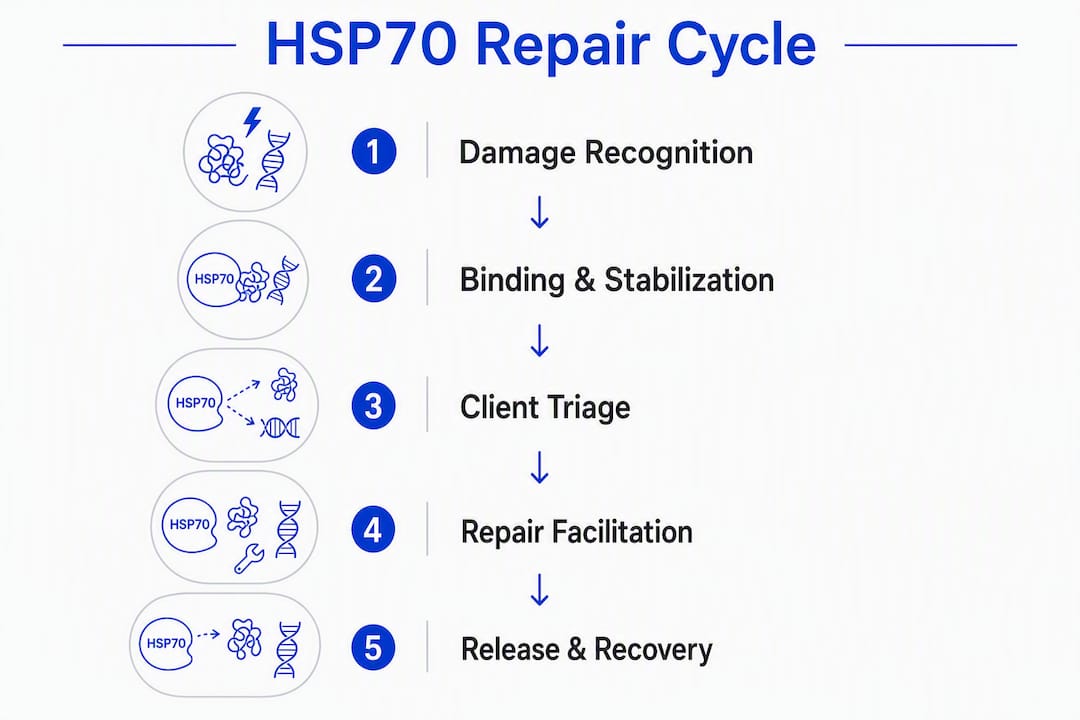
How does HSP70 repair or resolve damaged proteins?
The process begins with recognition, not random collision. A JDP surveys the proteome for exposed hydrophobic patches, the hallmark of misfolded or stress-damaged proteins, and delivers the client to HSP70 while simultaneously stimulating ATP hydrolysis. The resulting ADP-bound HSP70 grips the client tightly. What happens next depends entirely on which NEF and which downstream co-chaperones are present.
The four main outcomes, in order of increasing damage severity:
- Refolding: A NEF (Hsp110 or a BAG protein) exchanges ADP for ATP, opening the SBD and releasing the client into solution. If the client’s folding energy landscape is intact, it refolds spontaneously or with Hsp90 assistance. This is the preferred outcome for mildly denatured proteins.
- Holding: Under acute, overwhelming stress, HSP70 can bind clients without completing the ATPase cycle, effectively sequestering them until conditions improve. This prevents aggregation without committing to any fate.
- Disaggregation: Once aggregates form, metazoans use a three-component system: HSP70 plus a specialized JDP (DNAJB1 in humans) plus Hsp110 as NEF. This triad threads into aggregate surfaces and extracts polypeptides for refolding or degradation. Bacteria and yeast use Hsp100 disaggregases (ClpB/Hsp104) for the same job, but metazoans lost Hsp100 and rely entirely on this HSP70-centered machinery.
- Delivery for degradation: CHIP ubiquitinates the HSP70-bound client, flagging it for the 26S proteasome. BAG1 physically bridges HSP70 to the proteasome lid. For larger aggregates or when the proteasome is saturated, BAG3 redirects clients to chaperone-mediated autophagy (CMA) or macroautophagy.
The PMC mechanistic review describes HSP70 as a “binding platform” where the final client fate depends on the full interaction set assembled around it. The “multiple-socket” framing captures this well: HSP70 is the hub, and each co-chaperone is a socket that, when occupied, biases the outcome.
Experimental evidence for these steps comes from co-immunoprecipitation (co-IP) showing HSP70 in complex with misfolded clients, in vitro refolding assays using chemically denatured luciferase or citrate synthase as substrates, and proteasome-delivery assays tracking ubiquitinated client flux.

Pro Tip: To separate folding activity from degradation targeting in cell-based assays, use a CHIP knockout or CHIP-ΔTPR mutant as your degradation-null control alongside wild-type HSP70. Any client accumulation you see in the CHIP-null background that disappears when you add back CHIP is degradation-dependent, not folding-dependent. Pair this with a BAG1 knockdown to confirm proteasome routing versus autophagy.
How does HSP70 contribute to DNA repair beyond the cytoplasm?
The cytoplasmic folding role is well established. The nuclear role is less widely appreciated but mechanistically compelling, and it is where the role of HSP70 in repair becomes genuinely surprising.

Direct stimulation of APE1 in base excision repair
The clearest biochemical evidence comes from Mendez et al., who showed that HSP70 physically binds APE1 (also called HAP1 or APEX1) and accelerates its incision of abasic sites in BER. Critically, this stimulation is ATP-independent. HSP70 is not refolding APE1; it is acting as a structural scaffold that enhances APE1’s catalytic geometry. This distinction matters experimentally: ATPase inhibitors such as VER-155008 will not block the APE1 stimulation, so researchers who use them as a proxy for “all HSP70 activity” will miss this interaction entirely.
XRCC1/PARP1 complexing and single-strand break protection
Kotoglou et al. demonstrated in HeLa cells that HSP70 translocates to nuclei and nucleoli under heat stress, where it co-localizes and forms complexes with both XRCC1 and PARP1. XRCC1 is the scaffold protein that coordinates the BER/SSBR machinery, recruiting DNA polymerase β and DNA ligase III. PARP1 detects single-strand breaks and initiates the repair response by synthesizing poly(ADP-ribose) chains. HSP70 silencing in that model increased DNA strand breaks under heat stress, confirming a functional, not merely physical, contribution to repair.
Key mechanistic point: HSP70’s nuclear repair function is highly conditional on stress-induced translocation. Basal nuclear pools are low, so the repair-scaffolding activity is largely absent under normal growth conditions and becomes relevant specifically when damage accumulates.
Cross-pathway interactions beyond BER
HSP70’s reach extends into nucleotide excision repair (NER) and double-strand break (DSB) repair, though the evidence is less direct. The Springer review on heat shock proteins and DNA repair documents HSP70 family members as major components of heat-induced nuclear protein increases (HIENP), historically linked to altered DNA replication and repair kinetics after heat shock. For DSB repair, the connection runs partly through the broader HSP network: Hsp90 stabilizes RAD51 and BRCA1, and HSP70 feeds into this network by maintaining Hsp90 client loading. Inhibiting HSP70 therefore indirectly destabilizes homologous recombination (HR) factors, a mechanism that preclinical studies have exploited by combining HSP inhibitors with PARP inhibitors to sensitize tumor cells.
The linkinghub review frames this cross-talk explicitly: HSP70 serves as a scaffold connecting proteostasis and genome maintenance, and the two systems are not parallel but interdependent under stress.
What regulates HSP70’s repair activities at the molecular level?
Co-chaperone control
The co-chaperones described in the family overview section do not simply assist HSP70; they determine which repair-relevant function it performs. In the nucleus, the JDP landscape shifts. DNAJC2 (also called ZRF1/MPP11) associates with chromatin and has been linked to nucleotide excision repair. The BAG proteins show differential nuclear expression: BAG6 has a well-documented nuclear role in quality control of mislocalized proteins, and emerging evidence places it near DNA damage sites. CHIP’s nuclear presence is lower than its cytoplasmic abundance, but its activity at the nucleus can degrade repair factors that are themselves damaged, a function that becomes pathological when CHIP is overactive.
Post-translational modifications
| Modification |
Site |
Documented Effect on Repair-Relevant Function |
| Phosphorylation |
T495 |
Modulates G1/S checkpoint progression after DNA damage; alters interaction with repair co-factors |
| Phosphorylation |
— |
Affects ATPase activity and client release kinetics |
| Acetylation |
K71 |
Reduces ATPase activity; shifts balance toward client holding |
| Acetylation |
— |
Alters SBD conformation and client specificity |
| SUMOylation |
Multiple |
Promotes nuclear retention under stress |
The T495 phosphorylation site is the most repair-relevant. Work from eLife using kinase-hijacking strategies in pathogen models exposed T495 as a regulatory node: phosphorylation at this position affects G1/S progression after DNA damage, meaning it couples HSP70’s chaperone state to cell-cycle checkpoint control. Researchers studying repair kinetics who do not assay T495 phosphorylation status risk misinterpreting their results, particularly in experiments that span the G1/S boundary.
Nuclear localization dynamics
Under basal conditions, HSP70 is predominantly cytoplasmic. Heat shock, oxidative stress, and genotoxic agents drive rapid nuclear and nucleolar accumulation. The mechanism involves both passive diffusion of newly synthesized HSP70 (which lacks a classical NLS) and active import facilitated by importin interactions under stress conditions. Nucleolar accumulation is particularly pronounced and may reflect a role in ribosomal RNA quality control alongside DNA repair scaffolding. The low basal nuclear pool is a genuine constraint: repair-scaffolding activity is essentially stress-gated, which has implications for how you design experiments at physiological versus stress-mimicking temperatures.
Which experiments actually prove HSP70’s repair roles?
- Co-IP and pull-down: The standard entry point for mapping HSP70 interactions with APE1, XRCC1, and PARP1. Use nuclear fractions, not whole-cell lysates, to avoid cytoplasmic HSP70 swamping the signal. Include an RNase A treatment to rule out RNA-bridged interactions.
- In vitro APE1 incision assay: Incubate recombinant APE1 with a synthetic oligonucleotide containing a tetrahydrofuran (THF) abasic site analog, add purified HSP70, and measure nick frequency by denaturing PAGE. This directly tests stimulation without cellular complexity. Run parallel reactions with ATPase-dead HSP70 (K71A mutant) to confirm ATP independence.
- Comet assay (alkaline): Measures total single- and double-strand breaks at the single-cell level. Useful for quantifying DNA damage accumulation after HSP70 knockdown under heat or oxidative stress. Pair with a γH2AX foci count (immunofluorescence) to specifically track DSBs.
- Reporter-based repair assay: Plasmid-based BER or NER reporters transfected into HSP70-depleted cells give a functional readout of repair efficiency. The I-SceI-based DSB reporter (DR-GFP) can be adapted to test HR competence when HSP70 is manipulated.
- siRNA/CRISPR loss-of-function with rescue: Knock down HSPA1A specifically (not HSPA8/Hsc70) using isoform-specific siRNA or CRISPR guides, then rescue with siRNA-resistant wild-type or phosphomutant (T495A/T495D) constructs. This is the cleanest way to assign function to the inducible isoform.
- In vivo neuroprotection models: Rodent stroke models with HSP70 overexpression (adeno-associated virus delivery) or genetic knockout have been used to test whether the repair-scaffolding function translates to tissue protection. Readouts include lesion volume, comet assay on brain tissue, and XRCC1 foci quantification in neurons.
| Assay |
What It Proves |
Common Artifact |
| Co-IP (nuclear fraction) |
Physical interaction with repair proteins |
Cytoplasmic contamination; RNA bridges |
| APE1 incision assay |
Direct enzymatic stimulation |
Recombinant protein misfolding; buffer ATP contamination |
| Comet assay |
Cellular DNA strand break accumulation |
Lysis variability; electrophoresis inconsistency |
| γH2AX foci |
DSB formation and resolution kinetics |
Antibody cross-reactivity with H2AX variants |
| DR-GFP reporter |
HR efficiency |
Transfection efficiency variation |
| siRNA + rescue |
Isoform-specific function |
Off-target silencing; incomplete rescue |
Interpreting ATP dependence
This is where many studies go wrong. The classical HSP70 ATPase cycle is ATP-dependent, but the APE1 stimulation documented by Mendez et al. is not. If you add ATPase inhibitors and see no effect on APE1 activity, that does not mean HSP70 is uninvolved; it means the interaction is structural. Conversely, if you use ATP-depleted lysates and see loss of repair, you are likely disrupting the broader chaperone network, not specifically the APE1-scaffolding function. The cleanest approach is to use the K71A ATPase-dead mutant alongside wild-type HSP70 in both biochemical and cellular assays.
What do the highest-value primary studies actually show?
The mechanistic picture of HSP70 in DNA repair rests on a small number of well-designed primary studies, and understanding their specific experimental logic is more useful than citing them abstractly.
-
Mendez et al. (APE1 stimulation): Used purified recombinant HSP70 and APE1 in a cell-free incision assay. The stimulation was dose-dependent, reproducible across abasic site substrates, and ATP-independent. The study did not map the binding interface, which remains an open question. Replication in intact cells using nuclear fractionation and proximity ligation assay (PLA) would strengthen the structural model.
-
Kotoglou et al. (nuclear translocation and XRCC1/PARP1): Worked in HeLa cells under heat stress, combining immunofluorescence, subcellular fractionation, and co-IP to show translocation and complex formation. The functional consequence, increased DNA breaks after HSP70 silencing, was measured by comet assay. One limitation: the study used heat stress as the damage model, so whether the same complexes form after ionizing radiation or alkylating agents is not established.
-
eLife phosphorylation study (T495): Exploited pathogen kinase hijacking to identify endogenous regulatory sites. The T495 finding connects HSP70 phosphorylation to G1/S checkpoint control, but the direct mechanistic link to BER or SSBR enzyme activity has not been resolved. A phosphoproteomic time-course after defined DNA damage (e.g., methyl methanesulfonate treatment) would clarify whether T495 phosphorylation precedes or follows repair complex assembly.
-
Frontiers 2026 review: Integrates recent substrate specificity data and noncanonical HSP70 functions including RNA binding and anti-inflammatory roles. The Frontiers review is the most current synthesis of HSP70 functional scope and is worth reading alongside the Nature Reviews mechanistic paper.
Pro Tip: When reproducing the Kotoglou nuclear translocation result, use a cytoplasmic marker (e.g., GAPDH) and a nuclear marker (e.g., histone H3) in your fractionation western blots to confirm fraction purity before interpreting HSP70 redistribution. A contaminated nuclear fraction is the single most common reason this experiment fails to replicate.
What are the disease implications and therapeutic strategies?
Where HSP70’s repair roles matter clinically
Neuroprotection: Neurons are post-mitotic and cannot dilute damaged proteins or DNA through division. HSP70’s dual role in clearing proteotoxic aggregates and scaffolding BER/SSBR makes it particularly critical in stroke, where oxidative DNA damage and protein aggregation occur simultaneously. In vivo rodent stroke models with HSP70 overexpression show reduced lesion volume and improved neuronal survival, consistent with both repair functions contributing.
Cancer: Tumor cells frequently upregulate HSP70 to survive the proteotoxic and genotoxic stress of rapid proliferation. This upregulation also enhances DNA repair capacity, contributing to therapy resistance. The same HSP70 that helps a cancer cell survive chemotherapy-induced DNA damage is protecting its genome from the treatment.
Aging: Proteostasis capacity declines with age, partly because HSF1 activity drops and HSP70 induction becomes blunted. Reduced HSP70 means both more protein aggregation and less efficient BER, a combination that accelerates the genomic instability associated with aging tissues.
Modulation strategies and their trade-offs
- HSF1 activators (e.g., geranylgeranylacetone, celastrol): Upregulate the entire HSP70 network. Useful in neuroprotection models. Risk: broad transcriptional effects beyond HSP70; potential to protect tumor cells from therapy.
- Direct HSP70 inhibitors (e.g., VER-155008, MKT-077): Suppress ATPase activity or allosteric sites. Sensitize tumor cells to DNA-damaging agents by impairing repair scaffolding. Risk: normal tissue toxicity, particularly in neurons and cardiomyocytes that depend on constitutive HSP70 activity.
- CHIP (STUB1) modulation: Enhancing CHIP activity drives damaged proteins toward degradation, reducing the pool available for repair scaffolding. Inhibiting CHIP preserves repair-competent HSP70 complexes but risks accumulating misfolded proteins.
- BAG family targeting: BAG3 inhibition impairs autophagy-mediated clearance; BAG1 inhibition reduces proteasome delivery. Neither is repair-specific, but both alter the balance between repair and degradation of HSP70 clients.
- Combination strategies: Preclinical data support combining HSP network inhibitors with PARP inhibitors. Downregulating HSP70/Hsp90 reduces RAD51 and BRCA1 stability, impairing HR, while PARP inhibition blocks SSBR. The combination creates synthetic lethality in HR-deficient tumor models.
The key limitation across all strategies is selectivity. HSP70 is ubiquitous and multifunctional; any systemic intervention will affect proteostasis, DNA repair, and immune signaling simultaneously. Tissue-targeted delivery (e.g., AAV for neuronal overexpression, nanoparticle-encapsulated inhibitors for tumor targeting) is the direction the field is moving to address this.
What are the most important open questions in this field?
-
Isoform-specific nuclear repair roles: Does inducible Hsp70 (HSPA1A) perform functions in nuclear BER that Hsc70 (HSPA8) cannot substitute for, and if so, what structural feature accounts for the difference? Conditional HSPA1A knockout in specific cell types (neurons, proliferating epithelium) with HSPA8 intact would answer this directly.
-
How do specific PTMs route clients toward BER versus the proteasome? The T495 phosphorylation data suggest a cell-cycle coupling mechanism, but the downstream effectors are not mapped. A phosphoproteomic pulldown after defined damage, comparing T495A and T495D mutants, would identify the interactors that change.
-
Temporal sequence of HSP70 recruitment after damage: Does HSP70 arrive at damage sites before or after PARP1 activation? Live-cell imaging with tagged HSP70 and a PARP1 activity sensor (e.g., a PAR-binding domain fused to a fluorophore) could resolve recruitment order in real time.
-
NER and DSB repair: direct or indirect? The evidence for HSP70 in NER and HR is largely indirect, running through Hsp90 client stabilization. A proximity proteomics approach (BioID or TurboID fused to HSP70) in cells treated with UV (NER) or ionizing radiation (DSB) would map the nuclear interaction network under each damage type.
-
Disaggregation at chromatin: Can the metazoan HSP70/DNAJB1/Hsp110 disaggregation triad act on chromatin-associated protein aggregates, and does this contribute to repair factor accessibility? In vitro chromatin templates with defined aggregated repair proteins would test this.
-
Translational gap in neuroprotection: Most in vivo neuroprotection data use overexpression models. Pharmacological HSF1 activation in aged animals, where endogenous HSP70 induction is blunted, would better model the therapeutic scenario.
Priority experimental approaches: conditional tissue-specific knockouts (CRISPR in organoids), time-resolved proximity proteomics after defined damage, and single-molecule FRET to track HSP70 conformational states at repair foci. Funding agencies including the NIH National Institute of General Medical Sciences (NIGMS) and the National Cancer Institute (NCI) have active mechanisms supporting this type of mechanistic cell biology.
Key Takeaways
HSP70 functions as a stress-activated triage scaffold that directly stimulates BER enzymes, forms protective complexes with XRCC1 and PARP1 in the nucleus, and routes damaged proteins to refolding or degradation depending on which co-chaperones are present.
| Point |
Details |
| Dual repair roles |
HSP70 repairs damaged proteins via folding/disaggregation and supports DNA repair by scaffolding APE1, XRCC1, and PARP1. |
| ATP-independent DNA repair |
HSP70 stimulates APE1 endonuclease activity without ATPase cycling, so ATPase inhibitors alone cannot block this function. |
| Co-chaperone triage |
Hsp40/DnaJ, Hsp110, BAG family, and CHIP together determine whether a client is refolded, held, or degraded. |
| Phosphorylation at T495 |
T495 phosphorylation couples HSP70 activity to G1/S checkpoint control; assay this site when studying repair kinetics. |
| Stress-gated nuclear function |
HSP70 nuclear pools are low under basal conditions; repair scaffolding activity requires stress-induced translocation to engage XRCC1 and PARP1. |
Why HSP70’s cross-talk with genome maintenance deserves more attention
The field spent two decades treating HSP70 as a cytoplasmic folding machine with an occasional nuclear cameo. The mechanistic evidence now points to something more integrated: a protein whose co-chaperone context, phosphorylation state, and subcellular address collectively determine whether it is folding a misfolded kinase, routing an aggregated client to autophagy, or sitting on a BER complex accelerating APE1 incision.
What strikes me about this body of work is how much the therapeutic framing has lagged the basic science. Researchers designing HSP70 inhibitor studies for cancer often focus entirely on proteotoxic stress, ignoring that the same inhibition is simultaneously impairing BER scaffolding. That oversight probably explains some of the inconsistent sensitization results in the literature. Conversely, neuroprotection studies that upregulate HSP70 via HSF1 activators are likely getting benefit from both the proteostasis and the genome-maintenance arms, but the two contributions are rarely dissected.
For labs starting in this area: the most tractable entry point is the APE1 incision assay paired with isoform-specific siRNA. It is clean, reproducible, and directly tests the ATP-independent scaffolding function without the confounds of whole-cell overexpression. Add a T495 phosphomutant rescue and you have a two-experiment package that addresses both the biochemical mechanism and the regulatory switch. Collaborating with a structural biology group to map the HSP70-APE1 binding interface would be the logical next step, and it is the gap in the primary literature most likely to generate a high-impact finding in the next few years.
Useful sources for deeper follow-up
Researchers setting up HSP70 repair assays for the first time can also consult Synthrolab’s tissue repair peptide research protocol for practical experimental workflow guidance, and the Synthrolab test builder for structuring and documenting experimental designs in repair biology studies.
Recommended
 21+ verification