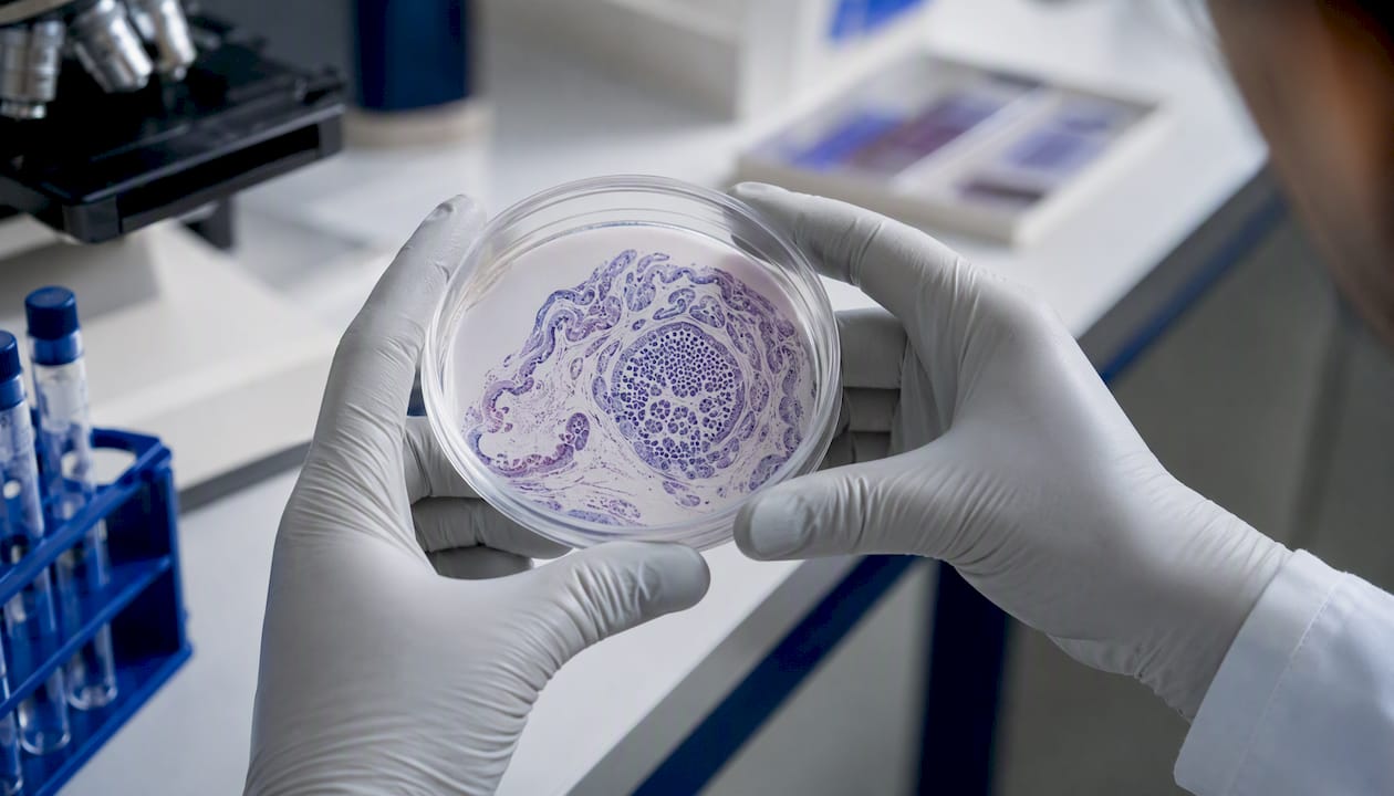
A stem cell niche is the local microenvironment that directly surrounds and controls a stem cell, determining whether it stays dormant, divides, or differentiates into a specialized cell type. Think of it less as a passive address and more as an active control system: the niche sends continuous molecular and physical instructions, and the stem cell responds in kind. That two-way relationship is what makes the niche so central to tissue biology.
The three primary functions the niche performs:
- Maintenance and quiescence: Keeps stem cells in a non-dividing, protected state during normal homeostasis, preventing premature exhaustion.
- Activation and regeneration: Releases stem cells from quiescence after tissue injury, triggering self-renewal or differentiation to repair damage.
- Homeostatic balance: Prevents both depletion (which causes tissue failure) and overproliferation (which risks malignant transformation).
Why does this matter beyond cell biology? Because niche dysfunction sits at the root of aging-related tissue decline, failed regeneration, and cancer. A niche that loses its supportive signals ages a tissue faster than the stem cells themselves do. One that sends aberrant proliferative cues can seed a tumor. Understanding the stem cell niche is, in practical terms, understanding how tissues stay healthy or fall apart.
Key Takeaways
The stem cell niche is a dynamic, two-way regulatory system where local microenvironment signals determine stem cell fate, and its dysfunction drives aging, failed regeneration, and cancer.
| Point |
Details |
| Niche definition |
A discrete local microenvironment that is both necessary and sufficient for stem cell maintenance, defined by anatomy and experimental function. |
| Core components |
Cellular (support cells, endothelium, immune cells) and acellular (ECM, oxygen tension, mechanical cues, metabolites) elements integrate to regulate stem cell output. |
| Key signalling pathways |
Wnt, Notch, BMP, Hedgehog, CXCL12, and mechanotransduction via integrins and YAP/TAZ are the dominant regulatory axes across most niches. |
| Experimental proof |
Niche identity requires convergent evidence: lineage tracing, transplantation, ablation, and ideally single-cell spatial profiling to confirm necessity and sufficiency. |
| Translational potential |
Niche-targeted drugs, biomaterial scaffolds, and peptide modulators are active research strategies, but tissue specificity and oncogenic risk remain the central obstacles. |
Table of Contents
What Is a Stem Cell Niche? Definition and the Niche Hypothesis
The stem cell niche is both an anatomical location and a functional unit. Anatomically, it is a discrete region within a tissue where stem cells reside in close contact with support cells and a specialized extracellular matrix. Functionally, it is defined by its ability to regulate stem cell behavior: a true niche must be shown experimentally to be both necessary and sufficient for stem cell maintenance.
The concept traces back to Raymond Schofield, who proposed in 1978 that hematopoietic stem cells depend on a specific cellular environment to retain their self-renewal capacity, a hypothesis that has since been widely supported by research. That idea, radical at the time, has since been validated across virtually every tissue studied. As Nature Reviews Molecular Cell Biology summarizes, niche features are conserved across diverse organisms and tissues: signals from support cells, cell adhesion, mechanical inputs, and spatial cues appear repeatedly, and niches form at discrete developmental times to regulate stem cell proliferation and fate.
Three criteria distinguish a true niche from a mere anatomical neighborhood. First, anatomic specificity: stem cells cluster in a defined region, not randomly throughout a tissue. Second, functional regulation: removing or altering niche components changes stem cell behavior in predictable ways. Third, experimental testability: the niche can be probed by ablation, transplantation, or genetic manipulation, and the results are reproducible. Displacement experiments confirm this directly: moving a stem cell away from its niche contact typically causes rapid loss of self-renewal capacity, which is about as clean a demonstration of niche dependence as biology offers.
What Are the Cellular and Acellular Components of a Niche?
The niche is not a single molecule or cell type. It is an integrated system of cellular neighbors and physical materials that together produce the regulatory environment a stem cell reads.
Cellular components include:
- Stromal support cells (fibroblasts, mesenchymal stromal cells): secrete growth factors, deposit ECM, and present membrane-bound ligands like Notch and Wnt pathway components.
- Endothelial cells lining nearby vasculature: supply oxygen, nutrients, and paracrine signals including CXCL12 and angiopoietins; vascular proximity is a defining feature of hematopoietic and neural niches.
- Neuronal inputs: sympathetic nerve fibers innervate several niches (notably the bone marrow) and modulate stem cell mobilization through adrenergic signaling.
- Immune cells: macrophages and regulatory T cells contribute anti-inflammatory signals and phagocytic clearance that protect stem cell integrity.
- Differentiated progeny: mature daughters of stem cells can feed back inhibitory signals (Notch ligands, BMPs) that limit further stem cell output, a classic negative feedback loop.
Acellular components are equally important and often underappreciated:
- Extracellular matrix (ECM): fibronectin, laminin, collagen isoforms, and heparan sulfate proteoglycans provide adhesion anchors and act as a reservoir for growth factors. Matrix architecture, not just composition, matters: fiber alignment and crosslinking density alter how cells sense and respond to the matrix.
- Immobilized versus soluble growth factors: factors tethered to the ECM (e.g., FGF2 bound to heparan sulfate) act locally and with higher effective concentration than their soluble counterparts.
- Oxygen tension: many niches are hypoxic (bone marrow perivascular zones, intestinal crypt bases), and low oxygen stabilizes HIF-1α, which supports quiescence and glycolytic metabolism.
- Metabolites and pH: lactate, succinate, and NAD+/NADH ratios influence epigenetic enzyme activity and thus gene expression in stem cells.
- Mechanical cues: substrate stiffness, shear stress from blood flow, and hydrostatic pressure all feed into stem cell fate decisions. Research published in Stem Cell Research & Therapy confirms that ECM stiffness, shear stress, and substrate topology shape stem cell differentiation by engaging mechanosensing cascades that interact directly with chemical signaling.
These components do not act in isolation. The niche integrates redundant and sometimes opposing cues: a quiescence-promoting BMP signal from one cell type may be counterbalanced by a proliferative Wnt signal from another. The net output depends on signal strength, receptor expression, and the stem cell’s own epigenetic state. That complexity is precisely why replicating a niche in a dish has proven so difficult.
Which Signalling Pathways Drive Niche–Stem Cell Communication?
Several conserved pathways carry the bulk of niche-to-stem-cell instructions. The table below maps each to its canonical role, typical niche source, and a tissue example.
| Pathway |
Role in stem cell regulation |
Typical niche source |
Canonical tissue example |
| Wnt/β-catenin |
Promotes self-renewal and proliferation |
Paneth cells, stromal fibroblasts |
Intestinal crypt stem cells |
| Notch |
Maintains progenitor identity; lateral inhibition |
Adjacent support cells, endothelium |
Intestinal and hematopoietic niches |
| BMP/TGF-β |
Promotes quiescence and differentiation |
Stromal cells, bone matrix |
Hair follicle bulge; bone marrow |
| Hedgehog (Hh) |
Regulates stem cell number and niche size |
Stromal mesenchyme |
Hair follicle, neural stem cells |
| CXCL12/CXCR4 |
Retention and homing of stem cells to niche |
Endothelial and CXCL12-abundant reticular cells |
Hematopoietic bone marrow niche |
| Cytokines (SCF, TPO, IL-3) |
Survival, proliferation, lineage bias |
Stromal and endothelial cells |
Hematopoietic stem cells |
| Integrin/FAK/Src |
Adhesion and mechanosensing |
ECM (fibronectin, laminin) |
Muscle satellite cells, HSCs |
| YAP/TAZ (mechanotransduction) |
Translates ECM stiffness into gene expression |
ECM stiffness gradient |
Mesenchymal and epithelial niches |
Beyond ligand-receptor signaling, mechanotransduction deserves its own emphasis. ECM stiffness activates integrins, which recruit focal adhesion kinase (FAK) and trigger downstream cascades that converge on YAP and TAZ transcriptional co-activators. Piezo1 and Piezo2 ion channels respond to membrane tension and shear. These mechanical routes are not parallel to chemical signaling: they intersect with Wnt, Hippo, and TGF-β pathways at multiple nodes, meaning a change in matrix stiffness can amplify or dampen a chemical signal without any change in ligand concentration.
Metabolic coupling adds another layer. Stem cells in hypoxic niches rely heavily on glycolysis rather than oxidative phosphorylation, and this metabolic state is not just a consequence of low oxygen but an active regulatory choice. The PGC-1α pathway links mitochondrial biogenesis to niche oxygen availability, and disrupting this coupling can push stem cells out of quiescence prematurely.
Extracellular vesicles and exosomes have emerged as a distinct communication channel. Niche cells package proteins, microRNAs, and mRNAs into vesicles that fuse with stem cell membranes, delivering regulatory cargo without direct cell contact. This mechanism extends niche influence beyond the immediate contact zone and may explain how systemic signals (circulating EVs from distant organs) modulate local stem cell behavior.
How Do Researchers Identify and Test Niches?
Establishing that a microenvironment is a functional niche, not just a neighborhood, requires multiple converging experimental strategies. Each method answers a different question, and each has real interpretive limits.
| Method |
What it shows |
Main limitation |
| Lineage tracing |
Tracks the progeny of a labeled stem cell over time; confirms self-renewal and multipotency in vivo |
Label leakage or mosaic expression can misidentify the traced population |
| Transplantation |
Tests whether a donor stem cell can reconstitute a tissue in a conditioned host; probes niche sufficiency |
Host conditioning (irradiation) alters the niche, so results reflect a perturbed environment |
| Niche cell ablation |
Removes specific support cells to test niche necessity; stem cell loss or activation follows |
Off-target effects; ablation rarely removes one cell type cleanly |
| In vivo live imaging (intravital microscopy) |
Visualizes stem cell behavior and niche interactions in real time |
Technically demanding; limited tissue depth; phototoxicity risk |
| Single-cell RNA-seq + spatial transcriptomics |
Maps cell types, signaling states, and spatial relationships at single-cell resolution |
Snapshot in time; dissociation artifacts; spatial methods still limited in resolution |
| Organoids and engineered systems |
Recapitulates niche-dependent self-organization in 3D; modular control of individual parameters |
Lacks vasculature, immune cells, and systemic inputs; may not reflect in vivo dynamics |
The hematopoietic system provides a clear case study where lineage tracing with inducible Cre systems under HSC-specific promoters (e.g., Scl-CreERT) suggests that single HSCs can give rise to all blood lineages over months, satisfying self-renewal and multipotency criteria. Transplantation into lethally irradiated hosts then tests whether the bone marrow niche is sufficient to support donor HSC engraftment, which it is, but only when the perivascular and endosteal niche architecture remains intact. When CXCL12-abundant reticular cells are ablated, HSCs mobilize into the blood and lose quiescence, demonstrating niche necessity.
Pro Tip: When interpreting transplantation or ablation results, always ask whether the experimental manipulation itself altered the niche. Irradiation, for example, damages endothelial cells and triggers inflammatory remodeling that would not occur in normal homeostasis. A stem cell that “fails to engraft” may be responding to a disrupted niche, not an intrinsic defect. Pair every perturbation experiment with niche-composition readouts (immunostaining, scRNA-seq of support cells) to separate stem cell behavior from niche artifact.
Array-based material experiments allow testing of hundreds of microenvironmental conditions simultaneously, which accelerates discovery about how biochemical and biophysical cues influence stem cells far beyond what serial single-condition experiments can achieve.
How Are Niches Organized in Specific Tissues?
General niche principles become concrete when mapped onto specific tissues. Five canonical examples illustrate how the same logic plays out in very different anatomical contexts.
Hematopoietic stem cell niche (bone marrow)
HSCs occupy two overlapping zones: the endosteal niche (near trabecular bone surfaces, rich in osteoblasts) and the perivascular niche (adjacent to sinusoidal endothelium and CXCL12-abundant reticular cells). Quiescence is maintained by CXCL12, SCF, TGF-β, and angiopoietin-1. Key markers include CD34, CD38 (low), Lin (negative), Sca-1, and c-Kit in mice; CD34+CD38lowCD90+CD45RA- in humans. The two zones are not mutually exclusive: the deepest quiescent HSCs tend to be perivascular but also close to endosteal surfaces in trabecular-rich regions.
Intestinal stem cell niche (crypt base)
Intestinal stem cells (ISCs) sit at the base of crypts, interleaved with Paneth cells. Paneth cells are the dominant niche support cell here: they secrete Wnt3, EGF, Notch ligands (DLL4), and the R-spondin co-receptor ligand that amplifies Wnt signaling. The result is a steep Wnt gradient from crypt base to villus tip that drives a proliferation-to-differentiation axis. ISC markers include Lgr5 (a Wnt target itself), Olfm4, and Ascl2. The intestinal niche is among the best-characterized, partly because Lgr5-EGFP mice allow direct visualization of niche-resident stem cells.
Hair follicle bulge niche
Hair follicle stem cells (HFSCs) reside in the bulge, a protected compartment in the outer root sheath. During the resting phase (telogen), BMP signals from the dermal papilla and adipocyte layer maintain quiescence. Wnt activation from the dermal papilla triggers anagen (growth phase) entry. The bulge is also home to melanocyte stem cells, which depend on adjacent keratinocytes for Notch-mediated maintenance. Key markers: CD34, Sox9, Lgr5 (in secondary hair germ), and Keratin 15.
Muscle satellite cell niche
Satellite cells sit between the basal lamina and the plasma membrane of muscle fibers, a position that provides both ECM contact (laminin via α7β1 integrin) and direct fiber contact (M-cadherin, Notch ligands from the fiber). At rest, satellite cells are quiescent; injury disrupts the basal lamina and releases HGF, FGF2, and IGF-1, triggering activation. Pax7 is the defining marker; MyoD marks activated cells committing to myogenic differentiation. For researchers studying this niche with peptide tools, Synthrolab’s muscle repair peptide guide covers experimental setups relevant to satellite cell activation.

Neural stem cell niche (subventricular zone and hippocampal dentate gyrus)
In the adult brain, neural stem cells (NSCs) in the subventricular zone (SVZ) contact both the ventricle (via a primary cilium exposed to CSF) and blood vessels. VEGF, CXCL12, and Notch signals from endothelial cells maintain NSC identity. In the dentate gyrus, NSCs sit in the subgranular zone between the granule cell layer and the hilus, receiving GABA from mature granule cells (which promotes quiescence) and glutamate from mossy fibers (which promotes activation). Sox2, Nestin, and GFAP mark quiescent NSCs; DCX marks newly born neurons.
How Do Niches Change Across Development, Aging, and Injury?
Niches are not static. They form, mature, and eventually deteriorate, and each phase has distinct consequences for stem cell behavior.
During embryonic development, “emerging niches” support progenitor populations that drive organogenesis. These early niches are often transient: they form at discrete developmental windows, provide the signals needed for organ patterning, and then remodel as the tissue matures. The adult maintenance niche that takes over is structurally and molecularly distinct, optimized for long-term homeostasis rather than rapid expansion. Research from PMC/NIH confirms that niche plasticity across development and adulthood is a conserved feature, with emerging niches supporting progenitors during organogenesis while adult niches maintain tissue-specific stem cells for homeostasis.
After injury, niches shift into a facultative activation mode. Inflammatory cytokines (TNF-α, IL-6, IL-1β) disrupt quiescence signals, ECM-degrading proteases (MMPs) release sequestered growth factors, and damaged cells send danger signals (DAMPs) that recruit immune cells. This coordinated response temporarily overrides the quiescence program to drive regeneration. The niche then re-establishes homeostatic signals once repair is complete, a process that can fail in chronic injury states.
Aging-related niche changes are well documented. In the bone marrow, osteoblast numbers decline, CXCL12 production drops, and inflammatory stromal cells accumulate. In muscle, satellite cell niches show increased TGF-β signaling and fibronectin deposition, which promotes fibrosis over regeneration. In the brain, NSC niches accumulate reactive astrocytes and lose pro-neurogenic VEGF signals. The common thread: aged niches shift from pro-regenerative to pro-inflammatory and pro-fibrotic states, and stem cells respond accordingly, not because they are intrinsically exhausted but because their instructions have changed.
How Does Niche Dysfunction Drive Disease?
When niche regulation fails, the consequences extend well beyond impaired regeneration. The niche can become an active driver of pathology.
Cancer stem cell niches are the clearest example. In acute myeloid leukemia (AML), malignant stem cells remodel the bone marrow niche to their own advantage: they suppress normal osteoblast function, expand CXCL12-producing stromal cells, and recruit immunosuppressive regulatory T cells. The result is a niche that preferentially supports leukemic stem cell survival while actively excluding normal HSCs. This niche remodeling is a major reason why residual leukemic stem cells survive chemotherapy: the niche provides physical protection (adhesion-mediated quiescence) and anti-apoptotic signals that chemotherapy cannot easily overcome.

In solid tumors, cancer-associated fibroblasts (CAFs) and tumor-associated macrophages (TAMs) construct a niche that supports cancer stemness, promotes epithelial-to-mesenchymal transition, and facilitates metastatic seeding at distant sites. ECM stiffening in tumors, driven by collagen crosslinking via lysyl oxidase (LOX), activates integrin-FAK-YAP signaling in cancer cells and reinforces a stem-like, therapy-resistant phenotype.
Fibrosis represents a different failure mode. In liver fibrosis, hepatic stellate cells activate into myofibroblasts that deposit excessive collagen, stiffening the ECM and disrupting the normal hepatic stem cell niche. The resulting environment promotes further stellate cell activation (a feed-forward loop) and impairs hepatocyte regeneration. Similar dynamics occur in pulmonary fibrosis and chronic kidney disease.
PMC/NIH research makes the mechanism explicit: loss of normal niche input may permit overproliferation of stem cells, predisposing to malignant transformation, while aberrant niche signals can actively drive tumorigenesis. The clinical implication is that niche-targeted therapies need to restore normal regulatory balance, not simply block one pathway, because the niche operates through redundant, interconnected signals.
How Can Researchers Engineer and Recreate Niches in the Lab?
Recreating a niche in vitro requires simultaneous control of parameters that natural tissues integrate automatically. The main engineered systems each capture part of that complexity.
Common engineered platforms:
- Organoids: 3D self-organizing cultures derived from stem cells embedded in Matrigel or defined hydrogels. They recapitulate crypt-villus architecture (intestinal organoids) or neural rosette organization, but lack vasculature, immune cells, and systemic inputs. Best for studying epithelial self-organization and Wnt/Notch pathway dynamics.
- Hydrogel-based ECM mimics: Polyethylene glycol (PEG), hyaluronic acid, or fibrin hydrogels with tunable stiffness and functionalized with adhesion peptides (RGD, IKVAV, YIGSR). Allow independent control of stiffness and ligand density, which is impossible in Matrigel. Annual Reviews research confirms that engineered material systems enable controlled investigation of biochemical and biophysical niche cues, revealing the importance of immobilized biochemical and solid-phase mechanical signals.
- Peptide-functionalized surfaces: 2D or 3D substrates presenting bioactive peptide sequences that mimic ECM binding domains or growth factor receptor ligands. Useful for high-throughput screening of adhesion and signaling cues.
- Microfluidic devices: Introduce flow, oxygen gradients, and compartmentalized co-culture. Particularly useful for modeling vascular niches and studying how shear stress modulates stem cell behavior.
Practical parameters to control:
- Stiffness: Match to the target tissue (brain: ~0.1–1 kPa; muscle: ~8–17 kPa; bone: >25 kPa). Stiffness alone can direct mesenchymal stem cell lineage choice without added soluble factors.
- Ligand density and spacing: RGD peptide density controls integrin clustering and downstream FAK activation; spacing below ~70 nm is required for efficient integrin engagement.
- Growth factor presentation: Immobilized EGF or FGF2 (tethered via PEG linkers) produces more sustained receptor activation than soluble delivery and better mimics in vivo ECM-bound factor presentation.
- Oxygenation: Use hypoxia chambers or oxygen-permeable membranes to maintain physiological oxygen levels (1–5% O₂ for most niches, not the 21% of ambient air).
- Spatial patterning: Photopatterning or bioprinting can create gradients of stiffness or ligand density that mimic the crypt-to-villus or endosteal-to-vascular gradients in vivo.
A critical practical point: tuning one parameter in isolation often produces misleading results because of signal redundancy and cross-talk. A hydrogel with the right stiffness but wrong ligand density, or correct ligand density but ambient oxygen, will not faithfully replicate niche biology. Researchers designing niche-mimetic systems should treat parameter sets as integrated conditions, not independent variables. Synthrolab’s tissue repair peptide research protocol provides practical guidance on incorporating peptide cues into engineered ECM systems for niche modulation studies.
What Are the Translational Strategies for Targeting Niches?
Bench discoveries about niche biology have generated several translational strategies, each at a different stage of development.
| Strategy |
Approach |
Status |
Main challenge |
| Niche-targeted small molecules |
Block aberrant niche signals (e.g., anti-TGF-β, LOX inhibitors for fibrosis) |
Preclinical to early clinical |
Systemic off-target effects; pathway redundancy |
| Biomaterial scaffolds |
Implantable ECM-mimetic scaffolds to support stem cell engraftment and function |
Preclinical (some clinical for bone/cartilage) |
Vascularization; immune response; long-term stability |
| Cell therapy + niche conditioning |
Co-transplant stem cells with support cells or pre-condition the host niche |
Preclinical to clinical (HSC transplant is established) |
Defining the right support cell complement; scalability |
| Exosome/EV-based delivery |
Use niche-derived EVs to deliver regulatory cargo to stem cells |
Early preclinical |
Targeting specificity; manufacturing scale; cargo loading |
| Peptide-based niche modulation |
Synthetic peptides mimicking ECM binding domains or growth factor receptor ligands |
Preclinical |
Stability in vivo; delivery route; dose optimization |
Ethical considerations are significant, as strategies that broadly activate stem cell proliferation carry inherent oncogenic risk, particularly in tissues with accumulated somatic mutations. Regulatory frameworks in the United States (FDA oversight of cell and gene therapies under 21 CFR Part 1271 and related guidance) require demonstration of safety alongside efficacy, and niche-modulating biologics face the same scrutiny as any cell therapy. Researchers should design studies with appropriate tumor-formation monitoring and limit systemic exposure where possible.
Peptide-based approaches occupy an interesting middle ground: they can be designed with short half-lives, local delivery routes, and reversible receptor interactions, which reduces but does not eliminate oncogenic risk. Synthrolab’s stem cell signaling peptide resources cover the experimental rationale for peptide modulators used in niche-relevant research contexts.
This article is general scientific information and does not constitute medical or clinical advice. Confirm regulatory requirements and safety protocols with qualified professionals before designing translational studies.
What Are the Open Questions and Methodological Limits?
The niche field has matured rapidly, but several fundamental questions remain genuinely unresolved.
Open scientific questions:
- How universal are niche components across tissues? Wnt and Notch appear repeatedly, but whether the same molecular logic applies to every stem cell population or whether tissue-specific wiring dominates is unclear.
- What is the precise mechanotransduction wiring? The integrin-FAK-YAP axis is well established, but how Piezo channels, nuclear lamins, and cytoskeletal tension integrate with classical ligand pathways at the molecular level is still being worked out.
- How do long-range systemic signals (circulating hormones, systemic inflammation, gut microbiome metabolites) intersect with local niche control? The boundary between “niche” and “systemic environment” is not sharp.
- Can aged niches be fully rejuvenated, or do accumulated epigenetic changes in support cells set a ceiling on restoration?
- What drives niche size and stem cell number homeostasis? Why do some niches support exactly one stem cell (Drosophila germline) while others support thousands (intestinal crypts)?
Methodological caveats:
- Species differences: Mouse niche biology does not always translate directly to humans. HSC niche organization in humans differs from mice in ways that matter for clinical translation.
- In vitro artifact risks: Organoids and hydrogel cultures lack vasculature, immune inputs, and systemic signals. Results from these systems should be validated in vivo before strong mechanistic claims are made.
- Lineage-tracing interpretation: Cre-based systems require careful validation of driver specificity. Leaky or mosaic expression can label non-stem cells and produce misleading fate maps.
- Single-cell data sampling bias: scRNA-seq captures a snapshot and is biased toward abundant cell types. Rare niche cells (e.g., CXCL12-abundant reticular cells) may be underrepresented without enrichment strategies.
Pro Tip: When making a claim that a specific cell type is a niche component, include at least two independent lines of evidence: one showing co-localization or contact (imaging, spatial transcriptomics) and one showing functional consequence of that cell type’s removal or alteration (ablation, conditional knockout). A single correlative finding is not sufficient to establish niche identity.
A Researcher’s Perspective on Studying the Niche
The most underappreciated problem in niche biology is not the complexity of the signals. It is the tendency to study one variable at a time in a system that integrates dozens simultaneously. A researcher who carefully tunes hydrogel stiffness but ignores oxygen tension, or who ablates one support cell type without checking what compensatory signals the remaining cells upregulate, will generate data that is technically rigorous and biologically misleading. The niche is not a list of components. It is a network, and networks have emergent properties that single-variable experiments cannot capture.
The practical implication: invest in multi-parameter experimental designs from the start. Spatial transcriptomics paired with functional perturbation, or engineered hydrogels where stiffness and ligand density are varied together in factorial designs, produce far more interpretable results than sequential single-variable studies. The UW Bioinspired Engineering approach of array-based high-throughput screening is a model worth adopting more broadly.
For researchers at the bench who want to model or modulate niche signals, Synthrolab’s cellular regeneration compound catalog includes research-grade peptides and compounds relevant to ECM interaction, growth factor receptor signaling, and metabolic coupling, each independently batch-tested with certificates of analysis. The goal is to give researchers the molecular tools to ask better niche questions, not to shortcut the experimental rigor the field demands.
This article is general information, not a substitute for advice from a qualified doctor. Consult a qualified healthcare professional about your own circumstances before acting on anything here.
Sources
Recommended
 21+ verification