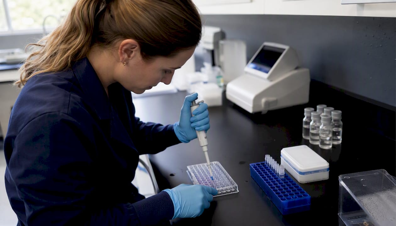
Cellular energy output is set by two proximal determinants: substrate flux through the TCA cycle and electron transport chain (ETC), and the structural integrity of the inner mitochondrial membrane (IMM) and its cardiolipin-dependent respiratory supercomplexes. Everything downstream of those two variables — ATP yield, membrane potential, proton leak — follows from them. For experimenters, that means three mechanistic levers are worth measuring before anything else:
- Substrate flux and coupling efficiency: track oxygen consumption in states 3 and 4, ATP production rate, and the P/O ratio
- Membrane health: measure mitochondrial membrane potential (Δψm) with TMRM or TMRE, and assess cardiolipin oxidation state by mass spectrometry or NAO staining
- Retrograde signalling: quantify AMPK phosphorylation, SIRT1 activity, and circulating or nuclear MOTS-c abundance alongside OXPHOS flux to connect metabolic signalling to ATP output
Elamipretide, the cardiolipin-binding aromatic-cationic peptide, and MOTS-c, a mitokine encoded in mitochondrial 12S rRNA, is the two most experimentally tractable entry points into those levers right now.
Table of Contents
The biochemical chain is linear but tightly regulated. Glucose enters glycolysis and yields pyruvate; fatty acids undergo beta-oxidation to acetyl-CoA. Both feed the TCA cycle, generating NADH and FADH2 as electron carriers. Those carriers donate electrons to Complexes I and II of the ETC, driving proton pumping across the IMM at Complexes I, III, and IV. The resulting proton motive force (Δp) powers ATP synthase (Complex V), converting ADP to ATP through oxidative phosphorylation (OXPHOS).
Fatty acid oxidation yields more ATP per carbon than glucose, but glucose oxidation is faster and more responsive to acute energy demand. The ratio matters for experimental design: substrate choice shifts measurable endpoints like the respiratory exchange ratio and state 3 oxygen consumption rate.
Two nutrient sensors govern substrate switching. AMPK (AMP-activated protein kinase) detects rising ADP/ATP ratios and activates catabolic pathways — fatty acid oxidation, glycolysis, mitochondrial biogenesis — while suppressing anabolic ATP consumers. SIRT1 senses the NAD+/NADH ratio and deacetylates PGC-1α and other transcriptional regulators to reprogram substrate preference. Both are experimentally accessible: AMPK phosphorylation at Thr172 by western blot, SIRT1 activity by fluorometric deacetylase assay, and NAD+ levels by enzymatic cycling assay or LC-MS.
Measurable endpoints for this layer of metabolism:
- Oxygen consumption rate (OCR) in states 3 and 4 via Seahorse XF or Clark-type electrode
- ATP production rate (luminescent ADP/ATP assay or luciferase-based)
- NAD+/NADH ratio (enzymatic cycling or LC-MS)
- ADP/ATP ratio (bioluminescent assay)
- P/O ratio (coupling efficiency from simultaneous O2 and ATP measurement)
Why does IMM structure determine how much ATP you actually get?
Substrate flux sets the ceiling; the IMM determines how much of that potential is captured as ATP rather than lost as heat. Cardiolipin, a dimeric phospholipid unique to the IMM, organizes respiratory complexes into supercomplexes (respirasomes) and stabilizes cristae curvature. When cardiolipin is oxidized or undergoes non-bilayer phase transitions, proton leak rises, supercomplex stability falls, and ATP output drops — independent of substrate availability.
Aromatic-cationic peptides including elamipretide (SS-31), SS-20, and HDAP2 bind cardiolipin directly, stabilize bilayer phases, reduce proton leak, and promote respiratory complex assembly. Elamipretide additionally interacts with ANT1 (adenine nucleotide translocase 1) and ETS supercomplex proteins, increasing ADP sensitivity and reducing ANT1-derived proton leak to improve in vivo ATP production. HDAP2 specifically addresses cardiolipin phase behavior: cardiolipin non-bilayer phases increase proton leakage and depolarization, and HDAP2 stabilizes bilayer cardiolipin to preserve membrane potential and maintain ATP output.
Key mechanistic insight: Optimizing mitochondrial ATP output is more about IMM structural integrity — cardiolipin stabilization, supercomplex assembly, and proton tightness — than broad ROS scavenging. Peptides that target cardiolipin address the proximal cause of energy loss, not a downstream symptom.
Practical metrics for this layer:
- State 4 respiration (proton leak index) via Seahorse or high-resolution respirometry
- Δψm by TMRM or TMRE fluorescence (plate reader or confocal)
- Cardiolipin oxidation by NAO (10-N-nonyl acridine orange) displacement or mass spectrometry
- Cardiolipin binding assays (liposome co-sedimentation or SPR with purified CL vesicles)
What do mitochondrial-derived peptides and micropeptides do to energy flux?
MDPs are a mechanistically distinct class. Rather than acting on membrane structure directly, they function as retrograde signals — mitochondria-to-nucleus messengers that reprogram nuclear gene expression to adjust cellular energy setpoints.
MOTS-c is a 16-amino-acid peptide encoded in the mitochondrial 12S rRNA gene that translocates to the nucleus under metabolic stress in an AMPK-dependent manner, where it regulates metabolism-associated transcription factors. The upstream mechanism: MOTS-c inhibits folate cycle and de novo purine biosynthesis, causing AICAR accumulation, which activates AMPK. Exogenous MOTS-c increases glucose uptake, modulates mitochondrial respiration, and improved running capacity and metabolic flexibility in animal models. MOTS-c levels decline with age, making age an important experimental variable. MOTS-c also engages SIRT1 and elevates NAD+ levels, connecting it to the same nutrient-sensing axis as AMPK.

Humanin and the SHLP family (SHLPs 1–6) are metaboloprotective MDPs that reduce apoptosis and modulate insulin signalling, though their direct ATP-output effects are less characterized than MOTS-c’s.
The newest mechanistic class is direct TCA modulators. APPM (ATP production-promoting micropeptide) is a 29-amino-acid peptide that binds the E1 subunit of OGDH (oxoglutarate dehydrogenase), increasing OGDHc activity to raise NADH and ATP production in vivo. Unlike MDPs, APPM acts at the enzyme level rather than through transcriptional reprogramming — a direct dial on TCA flux.
Mechanistic bullets for this section:
- MOTS-c: inhibits folate/purine biosynthesis → AICAR ↑ → AMPK activation → nuclear translocation → transcriptional reprogramming
- APPM: binds OGDH E1 subunit → OGDHc activity ↑ → NADH ↑ → ATP ↑
- Humanin/SHLPs: metaboloprotective, anti-apoptotic, insulin-sensitizing; measure via plasma ELISA and cell viability assays
Experimental readouts: nuclear MOTS-c localization by immunofluorescence or nuclear fractionation western blot; circulating MOTS-c by plasma ELISA; OGDH activity by spectrophotometric assay (NADH production rate) after APPM treatment; AMPK Thr172 phosphorylation as a downstream confirmation.
Pair real-time respirometry with endpoint ATP assays and Δψm measurement. That combination gives you flux (OCR), coupling efficiency (P/O), and membrane integrity in the same experiment. Seahorse XF Analyzer covers most cell and isolated mitochondria work; high-resolution respirometry (Oroboros O2k) is better for tissue homogenates and complex substrate titrations.
| Assay |
Readout |
Sample Type |
Key Caveat |
| Seahorse XF respirometry |
OCR, ECAR, state 3/4 |
Intact cells, isolated mitochondria |
Normalize to cell number or CS activity |
| Clark-type O2 electrode |
State 3/4 OCR, P/O ratio |
Isolated mitochondria, tissue |
Requires fresh isolation; buffer O2 calibration |
| Luciferase ATP assay |
Intracellular ATP |
Cells, tissue lysate |
Lysis method affects yield; use same protocol across conditions |
| TMRM/TMRE fluorescence |
Δψm |
Intact cells |
Quench mode vs. redistribution mode; validate with FCCP |
| NAO / mass spec |
Cardiolipin oxidation state |
Cells, isolated mitochondria |
NAO is semi-quantitative; MS gives oxidized species |
| AMPK Thr172 western |
AMPK activation |
Cell lysate, tissue |
Phosphatase inhibitors required in lysis buffer |
| Nuclear fractionation + western |
MOTS-c nuclear localization |
Cells |
Verify fraction purity with lamin A/C and GAPDH markers |
Design checklist for any peptide metabolism experiment:
- Include a vehicle control matched to peptide solvent (DMSO, sterile water, or BSA-conjugated for fatty acids)
- Run a full dose-response: in vitro typically 0.1–10 µM; in vivo reference ranges from published literature (e.g., MOTS-c mouse studies use 5–15 mg·kg−1 i.p.)
- Verify peptide cellular uptake and mitochondrial targeting before interpreting functional readouts
- Select model age deliberately — MOTS-c effects differ between young and aged animals
- Include a time course: acute (1–6 h) vs. chronic (24–72 h or multi-day in vivo) responses differ mechanistically
- Normalize respirometry data to citrate synthase (CS) activity or total protein, not just cell count
Pro Tip: Verify peptide mitochondrial targeting with a fluorescently tagged analog (e.g., FITC-conjugated peptide + MitoTracker co-localization) or subcellular fractionation before running full functional assays. Extracellular peptide effects can mimic intracellular ones and produce false-positive respirometry results.
What are the regulatory and translational limits of this research?
Most research peptides, including MOTS-c, elamipretide analogs, SS-20, HDAP2, and APPM, are sold as research-use-only (RUO) compounds. That designation means they are not approved for human therapeutic use and cannot be marketed or used with clinical efficacy claims. Any experiment designed to support an IND or clinical application requires a separate regulatory pathway through the FDA.
Practical regulatory cues for U.S. labs:
- Animal dosing studies: require IACUC approval; protocol must specify species, route of administration, dose range, and humane endpoints
- Human sample collection (blood, tissue for ex vivo assays): requires IRB approval even when no intervention is applied to the human subject
- RUO material transfer: document chain of custody; some peptides may require material transfer agreements (MTAs) between institutions
- Controlled substances: most metabolic peptides are not scheduled, but verify with your institution’s biosafety and compliance office before ordering
Regulatory caution: Do not make clinical efficacy or therapeutic claims based on RUO peptide experiments. Results from cell culture or animal models require independent validation and formal regulatory review before any clinical translation. Follow your institution’s biosafety, IACUC, and IRB requirements for every study design.
Translational limitations worth flagging explicitly:
- Species differences in MDP expression and receptor pharmacology can make rodent results poorly predictive of human outcomes
- Peptide cellular uptake varies by cell type, passage number, and membrane composition — always verify uptake in your specific model
- MOTS-c plasma levels decline with age, so aged animal cohorts will show different baseline and treatment responses than young animals
- MOTS-c effects can be tissue-specific and may be attenuated in severe mtDNA mutation models; validate your model’s mitochondrial genotype before scaling up
- APPM and HDAP2 are recently characterized; dose-response and off-target toxicity data in complex in vivo models remain limited
Key Takeaways
Cellular ATP output is determined by substrate flux through the TCA/ETC and by IMM cardiolipin integrity — measure both layers, not just one, to get a complete mechanistic picture.
| Point |
Details |
| IMM integrity is proximal |
Cardiolipin stability and supercomplex assembly are more direct ATP determinants than ROS levels; prioritize Δψm and proton leak assays. |
| Pair respirometry with ATP assays |
Seahorse OCR alone misses coupling efficiency; add a luciferase ATP assay and TMRM Δψm to complete the readout. |
| Verify peptide uptake first |
Confirm mitochondrial targeting by fluorescent co-localization or fractionation before interpreting any functional data. |
| Age is an experimental variable |
MOTS-c levels decline with age; always report cohort age and include age-matched controls in metabolic peptide studies. |
| Synthrolab for QC-backed reagents |
Synthrolab supplies batch-tested MOTS-c and metabolic modulation compounds with COAs, supporting incoming QC verification. |
The part most researchers skip
When designing metabolism experiments, the first question I ask is whether the model’s baseline mitochondrial function is actually characterized — not assumed. Most protocols jump straight to peptide treatment without establishing whether the cells or animals have the mitochondrial dysfunction the intervention is supposed to address. That single omission generates more irreproducible data than any assay choice.
Two other things get underweighted. First, normalization: OCR data normalized to cell count can be misleading when the intervention itself changes cell size or mitochondrial mass. Citrate synthase activity is a more stable denominator and takes 20 minutes to run. Second, the uptake verification step described above is treated as optional by most labs. It is not. A peptide sitting in the culture medium can activate surface receptors and produce OCR changes that look identical to mitochondrial targeting. Orthogonal assays — fractionation, fluorescent co-localization, or a cell-impermeable analog control — are what separate a mechanism from an artifact.
Share your COAs and raw assay files in supplementary data or a repository like Figshare. Reproducibility in this field depends on it.

Researchers investigating how metabolism affects energy levels at the cellular level need reagents they can trust before the experiment starts, not after a failed run.
Synthrolab supplies research-grade MOTS-c, elamipretide analogs, and related metabolic modulation compounds with batch-specific COAs that include HPLC chromatograms, MS confirmation, purity data, and endotoxin results. Every product ships lyophilized with documented storage and reconstitution guidance. COA access is available before purchase at synthrolab.com/coa, so you can run incoming QC verification against the vendor data from day one. All compounds are sold for research use only. Contact Synthrolab’s technical support team for experimental guidance on dosing, reconstitution, and assay compatibility before placing your first order.
Useful sources
Recommended
 21+ verification