LL-37 is the sole human cathelicidin, a 37-amino-acid cationic peptide that combines broad-spectrum antimicrobial activity with powerful, concentration-dependent immunomodulatory effects. Understanding it means holding two ideas at once: it kills bacteria by tearing apart membranes, and it shapes immune responses in ways that are genuinely context-dependent, sometimes protective, sometimes pathological.
Three things every researcher should know before designing an LL-37 experiment:
- Core mechanisms: direct membrane permeabilization, LPS/LTA neutralization, and pleiotropic immunomodulation through receptors including FPR2 and P2X7.
- Critical experimental caveats: LL-37 degrades rapidly in biological fluids, and cytotoxic effects appear in the 1–10 μM range, making concentration control non-negotiable.
- Sourcing: research-grade peptide with a verified Certificate of Analysis (CoA), confirmed purity by LC-MS, and a documented endotoxin value is the minimum acceptable standard. Synthrolab supplies independently batch-tested LL-37 for exactly this purpose.
Key Takeaways
LL-37 is the only human cathelicidin, and its concentration-dependent duality between antimicrobial killing and immunomodulation makes experimental rigor, particularly verified purity, endotoxin documentation, and full concentration-response data, the foundation of reproducible research.
| Point |
Details |
| Sequence and processing |
LL-37 (LLGDFFRKSKEKIGKEFKRIVQRIKDFLRNLVPRTES) is cleaved from hCAP18 by tissue-specific proteases; fragment identity must be confirmed in every experiment. |
| Concentration-dependent effects |
Cytotoxic effects appear in the 1–10 μM range; sub-μM concentrations tend toward anti-inflammatory activity, making full concentration-response curves mandatory. |
| Endotoxin control is non-negotiable |
LPS contamination in peptide preparations confounds all immune readouts; CoA must document an LAL-confirmed endotoxin value before use in cell-based assays. |
| Disease context shapes outcome |
LL-37 is protective in infection but pathological when overexpressed in rosacea, psoriasis, and certain cancers; in vivo context determines which effect dominates. |
| Synthrolab for sourcing |
Synthrolab provides independently batch-tested LL-37 with LC-MS identity, HPLC purity, and endotoxin documentation for laboratory research use. |
Table of Contents
What is the LL-37 peptide, and where does it come from?
LL-37 is derived from the precursor protein hCAP18, encoded by the CAMP gene on chromosome 3p21.3. The mature 37-residue sequence is:
LLGDFFRKSKEKIGKEFKRIVQRIKDFLRNLVPRTES
The name itself is structural: two leucines at the N-terminus, 37 residues total. Extracellular serine proteases, primarily kallikreins in the skin and proteinase 3 in neutrophils, cleave hCAP18 to release the active peptide. That processing step is not trivial. Different proteases generate different fragments with distinct activity profiles, and tissue expression and rapid local turnover mean systemic plasma concentrations stay low while local concentrations at infection or inflammation sites can climb orders of magnitude higher.
Amphipathic alpha-helical structure drives function
In aqueous solution, LL-37 is largely disordered. Contact with bacterial membranes or membrane-mimicking environments triggers folding into an amphipathic alpha helix spanning roughly residues 2–31. The helix positions a hydrophobic face against lipid acyl chains and a cationic face toward negatively charged phospholipid head groups. Net positive charge (approximately +6 at physiological pH) drives initial electrostatic attraction to bacterial membranes, which carry a net negative surface charge. No disulfide bonds constrain the structure, which is unusual among antimicrobial peptides and contributes to the peptide’s conformational flexibility.
Fragments used in research
Full-length LL-37 is not always the right tool. Several naturally occurring and synthetic fragments appear regularly in the literature:
| Fragment |
Residues |
Notes |
| KR-12 |
18–28 |
Shortest fragment retaining antimicrobial activity; lower host-cell cytotoxicity |
| GF-17 |
17–28 |
Retains strong membrane activity; studied for anti-biofilm properties |
| KR-20 |
18–37 |
Intermediate potency and toxicity profile |
| RK-31 |
7–37 |
Near full-length; similar activity to LL-37 |
Structural studies confirm that helix amphipathicity, rather than sequence length alone, determines antimicrobial potency, and that shorter fragments like KR-12 trade some potency for a meaningfully better cytotoxicity profile. Fragment selection is therefore an experimental design decision, not just a procurement one.
Statistic callout: LL-37’s molecular weight is approximately 4.5 kDa, and its net charge of approximately +6 at physiological pH places it firmly in the cationic antimicrobial peptide class, where electrostatic attraction to bacterial membranes is the first step in killing.
How does LL-37 kill microbes directly?
The 2025 Springer Nature review consolidates LL-37’s direct bactericidal actions into three overlapping mechanisms: membrane permeabilization, endotoxin neutralization, and biofilm disruption. Each operates at different concentration thresholds and against different microbial targets.
LL-37 inserts its hydrophobic face into bacterial lipid bilayers and disrupts membrane integrity through toroidal pore formation or a carpet-like detergent mechanism, depending on lipid composition and local peptide concentration. The result is ion leakage, loss of membrane potential, and ultimately cell lysis. This mechanism is concentration-dependent: sub-MIC concentrations may alter membrane fluidity without causing frank lysis, while concentrations above the MIC produce rapid killing.
LPS and LTA neutralization
Gram-negative bacteria release lipopolysaccharide (LPS) during infection, which drives TLR4-mediated inflammatory cascades. LL-37 binds LPS with high affinity through electrostatic and hydrophobic interactions, neutralizing its immunostimulatory activity. A parallel interaction with lipoteichoic acid (LTA) from Gram-positive organisms produces similar dampening of TLR2 signaling. This makes LL-37 simultaneously bactericidal and anti-endotoxin, a combination that is pharmacologically attractive but experimentally confounding: any immune readout in an LL-37 experiment must account for the peptide’s capacity to mask LPS activity in the assay medium.
Anti-biofilm activity
LL-37 disrupts established biofilm matrices and inhibits biofilm formation at sub-MIC concentrations. The mechanism involves both direct membrane disruption of sessile bacteria and interference with quorum-sensing signals. Synergy with conventional antibiotics has been reported in several organisms, though the magnitude varies considerably by species and biofilm maturity.
Activity spectrum and assay considerations
| Target |
Relative activity |
Key notes |
| Gram-negative bacteria |
Moderate to high |
LPS binding adds anti-endotoxin effect |
| Gram-positive bacteria |
Moderate |
LTA neutralization; variable MIC by species |
| Fungi |
Low to moderate |
Candida spp. most studied |
| Enveloped viruses |
Moderate |
Membrane disruption mechanism |
| Biofilms |
Moderate |
Sub-MIC activity; synergy with antibiotics |
Assay pitfalls for cationic peptides: LL-37 adsorbs to standard polystyrene plates, which artificially elevates apparent MIC values. Use low-bind polypropylene tubes and plates. High-salt buffers (above physiological ionic strength) reduce electrostatic interactions and suppress activity, so MIC broth microdilution should use cation-adjusted Mueller-Hinton broth at standard ionic strength. Time-kill assays should include a neutralizing agent at the sampling step to stop peptide activity and prevent carry-over killing artifacts.
How does LL-37 modulate immune responses?
Direct killing is only part of the story. LL-37’s immunomodulatory activities are broader, and in many clinical contexts, more consequential. The 2025 review documents NET stimulation, chemotaxis, and macrophage phenotype modulation as primary immunological outputs.
Key immunomodulatory functions:
- Chemotaxis: LL-37 recruits neutrophils, monocytes, and mast cells through FPR2 (formyl peptide receptor 2) binding, establishing a chemotactic gradient at infection sites.
- NET formation: LL-37 stimulates neutrophil extracellular trap (NET) release, which can trap and kill bacteria but also contributes to sterile inflammation and autoimmune pathology when dysregulated.
- Macrophage phenotype modulation: LL-37 shifts macrophage responses in a concentration- and context-dependent manner, promoting M1-like pro-inflammatory activation at high concentrations and supporting M2-like wound-healing phenotypes at lower concentrations in some models.
- Wound healing and angiogenesis: LL-37 activates epidermal growth factor receptor (EGFR) transactivation and promotes keratinocyte migration and proliferation, contributing to skin barrier repair.
- Type I interferon induction: LL-37 complexes with self-DNA or RNA, enabling uptake by plasmacytoid dendritic cells and activation of TLR7/TLR9 and STING pathways, driving type I IFN production. This mechanism is central to its role in autoimmune conditions including psoriasis and lupus.
Receptor interactions and downstream signaling
Beyond FPR2, LL-37 engages P2X7 purinergic receptors (promoting IL-1β release via NLRP3 inflammasome activation) and transactivates EGFR through metalloprotease-dependent shedding of EGF-like ligands. Downstream signaling cascades include MAPK/ERK, PI3K/Akt, and NF-κB pathways, which collectively regulate cytokine production, cell survival, and proliferation.
Concentration matters enormously here. At sub-μM concentrations, LL-37 tends to be anti-inflammatory, partly through LPS neutralization and partly through FPR2-mediated resolution signaling. At sufficiently elevated concentrations, pro-inflammatory outputs dominate. Endotoxin contamination in the peptide preparation shifts this balance unpredictably, which is why a documented endotoxin value in the CoA is not optional.
Cytotoxicity toward host cells can serve a biological purpose: LL-37 preferentially targets infected or apoptotic host cells, potentially limiting pathogen spread by eliminating cellular reservoirs. This nuance matters when interpreting in vitro cytotoxicity data, where the same concentration that kills bacteria may also reduce cell viability in ways that are physiologically meaningful rather than purely toxic.
Recommended signaling readouts: phospho-ERK1/2, phospho-p38, phospho-NF-κB p65, IL-6, IL-8/CXCL8, TNF-α, and type I IFN (IFN-α/β) by multiplex ELISA or Luminex. Transcriptomic endpoints (RNA-seq or NanoString) provide pathway-level resolution that single-cytokine panels miss.
Where does LL-37 drive disease, and where does it protect?
LL-37’s dual nature, protective at physiological concentrations and pathological when overexpressed or aberrantly processed, plays out differently across tissues.
Skin: rosacea and psoriasis
Elevated local cathelicidin production and altered proteolytic processing are directly implicated in rosacea pathogenesis. In affected skin, kallikrein 5 overactivity generates LL-37 fragments with enhanced inflammatory signaling relative to the full-length peptide, driving vasodilation, neutrophil recruitment, and the characteristic erythema. Standardizing which LL-37 fragment is being studied matters clinically: the rosacea-relevant fragments differ from those generated in healthy skin.
In psoriasis, LL-37 complexes with self-DNA released from dying keratinocytes. These complexes activate plasmacytoid dendritic cells via TLR9, initiating the type I IFN cascade that drives the psoriatic inflammatory loop. Local LL-37 concentrations in psoriatic plaques are elevated compared to normal skin, which is relevant when extrapolating in vitro data to disease physiology.
Respiratory mucosa and periodontitis
LL-37 is constitutively expressed in airway epithelium and is upregulated during pulmonary infection. It contributes to mucosal defense against Pseudomonas aeruginosa and other respiratory pathogens, though some organisms have evolved resistance mechanisms (discussed below). In the oral cavity, LL-37 in gingival crevicular fluid contributes to periodontal defense, and reduced expression has been associated with increased susceptibility to periodontitis.
Neuroinflammation
Microglial cells express and respond to LL-37. In neuroinflammatory contexts, LL-37 can activate microglia toward a pro-inflammatory phenotype, contributing to neuronal damage in conditions including Alzheimer’s disease, where LL-37 has also been studied for its anti-amyloid properties. The anti-amyloid activity, documented in structural studies, involves direct interaction with amyloid fibrils, though whether this is protective or pathological in vivo remains an open question.
Cancer: a genuinely dual role
LL-37’s cancer biology resists simple characterization. High-quality reviews document both pro- and anti-tumor effects depending on tumor type and microenvironment. In ovarian and lung cancers, LL-37 promotes tumor cell proliferation, angiogenesis, and metastasis through EGFR and Wnt pathway activation. In colorectal and gastric cancers, evidence points toward anti-tumor activity, including direct cytotoxicity toward cancer cells and immune activation. The tumor microenvironment, particularly its immunosuppressive or immunostimulatory character, appears to determine which effect dominates.
Clinical caveat: cytotoxic effects observed at μM concentrations in vitro do not translate directly to systemic therapeutic safety. Local tissue concentrations at sites of inflammation can reach μM levels naturally, but systemic administration at those concentrations carries genuine toxicity risk. Never extrapolate in vitro MIC or cytotoxicity data to therapeutic dosing without pharmacokinetic and toxicokinetic modeling.
What are the realistic therapeutic prospects for LL-37?
Native LL-37 faces three fundamental obstacles as a therapeutic: rapid proteolytic degradation in biological fluids, concentration-dependent cytotoxicity to host cells, and a pro-inflammatory risk at high local concentrations. None of these is insurmountable, but each requires deliberate engineering.
Delivery approaches under investigation:
- Topical formulations for wound healing and dermatologic conditions, leveraging local delivery to reduce systemic exposure.
- Inhaled aerosols for pulmonary infections, particularly against antibiotic-resistant P. aeruginosa.
- Nanoparticle carriers (liposomes, polymeric nanoparticles) that protect the peptide from proteolysis and enable controlled release.
- Hydrogel matrices for sustained local delivery in wound or periodontal applications.
Modification strategies and trade-offs:
- D-amino acid substitution: increases protease resistance substantially; may reduce immunogenicity but can alter receptor binding specificity.
- Cyclization: improves stability and can enhance membrane selectivity; synthesis is more complex.
- PEGylation: extends half-life and reduces immunogenicity; often reduces antimicrobial potency.
- Lipidation: enhances membrane affinity and self-assembly; increases cytotoxicity risk.
- Fragment use (KR-12, GF-17): lower cytotoxicity than full-length LL-37 while retaining antimicrobial activity; preferred for many therapeutic applications.
Preclinical safety checklist:
- Local cytotoxicity assays at the intended delivery site (keratinocytes, airway epithelial cells, or relevant primary cells).
- Cytokine storm risk assessment: measure IL-6, TNF-α, and IFN-γ in relevant immune cell co-culture models.
- Off-target membrane damage: erythrocyte hemolysis assay as a minimum screen.
- Protease stability profiling in relevant biological fluids (serum, bronchoalveolar lavage, wound exudate).
- Endotoxin-free preparation confirmed by LAL assay before any in vivo dosing.
How should you source, handle, and dose LL-37 in experiments?
Getting the peptide right before the experiment starts is where most reproducibility problems originate. The following checklist reflects minimum acceptable standards for LL-37 research.
Vendor selection and product acceptance testing
- Certificate of Analysis (CoA): confirm it covers the specific batch you receive, not a representative lot.
- Peptide identity: LC-MS confirmation of the correct molecular mass (approximately 4.5 kDa for full-length LL-37) and sequence.
- Purity: HPLC purity of ≥95% is the standard for functional assays; lower purity introduces unknown fragment contaminants that confound immunological readouts.
- Endotoxin: the CoA must state an endotoxin value in EU/mg or EU/µg, confirmed by LAL or recombinant Factor C assay. For immune cell assays, endotoxin below 1 EU/mg is a common threshold, though some assays require lower.
- Storage temperature: lyophilized LL-37 is stable at -20°C; reconstituted aliquots should be stored at -80°C and used within one freeze-thaw cycle where possible.
- Fragment annotation: confirm whether you are receiving full-length LL-37 or a specific fragment; the CoA should state the exact sequence.
Synthrolab provides independently batch-tested research-grade peptides with CoA documentation covering LC-MS identity, HPLC purity, and endotoxin values, along with reconstitution guidance and lab-grade consumables. For researchers sourcing LL-37, verifying CoA and storage conditions before committing to a batch is the single most impactful quality step.
Reconstitution and handling
Reconstitute lyophilized LL-37 in sterile, endotoxin-free water or 0.1% acetic acid (for poorly soluble batches), then dilute to working concentration in the appropriate assay buffer. Use low-bind polypropylene tubes throughout; standard polystyrene adsorbs cationic peptides and reduces effective concentration by a measurable margin. Endotoxin-free reconstitution solutions are not optional when immune readouts are the endpoint.

Pro Tip: Prepare a fresh dilution series for each experiment rather than storing working stocks. LL-37 activity declines with repeated freeze-thaw cycles, and adsorption to tube walls at low concentrations can produce apparent concentration-response relationships that are actually artifacts of surface binding.
Recommended concentration ranges and controls
Antimicrobial assays typically use LL-37 over a range of micromolar concentrations for MIC determination, though exact MICs vary by organism, growth phase, and media composition. Immunological assays commonly use 0.1–10 μM, with cytotoxicity becoming a significant confounder above approximately 5 μM in most primary cell systems.
Required controls for every LL-37 experiment:
- Vehicle control: matched solvent at equivalent volume.
- Scrambled peptide control: same amino acid composition, randomized sequence, to distinguish sequence-specific from physicochemical effects.
- Heat-inactivated peptide: confirms activity requires intact secondary structure.
- Endotoxin spike control: LPS at a concentration matching the endotoxin value in the CoA, to confirm that any immune response is peptide-mediated, not endotoxin-driven.
- Full concentration-response curve: never report a single-concentration result as definitive; LL-37’s concentration-dependent duality makes this non-negotiable.
Which assays and readouts work best for studying LL-37?
Functional antimicrobial assays
MIC broth microdilution is the standard for minimum inhibitory concentration. Use cation-adjusted Mueller-Hinton broth, low-bind plates, and include a peptide-free growth control and a sterility control. Read at 18–24 hours. Time-kill assays track colony-forming units over time at a fixed peptide concentration (typically 1× and 4× MIC) and reveal killing kinetics that MIC alone cannot capture.
MBEC (minimum biofilm eradication concentration) assays use the Calgary Biofilm Device or equivalent to quantify the concentration required to eradicate established biofilms. These concentrations are typically 10–1,000-fold higher than planktonic MICs, which has direct implications for therapeutic dosing calculations.
SYTOX Green or propidium iodide (PI) membrane integrity assays provide a rapid fluorescence-based readout of membrane permeabilization kinetics, useful for mechanistic studies and fragment comparisons.
Analytical quantitation methods
| Method |
Primary readout |
Typical sample |
Major pitfall |
| ELISA |
LL-37 concentration |
Plasma, BAL, wound fluid |
Antibody cross-reactivity with hCAP18 and fragments |
| LC-MS/MS |
Peptide mass and sequence |
Any biological matrix |
Requires method development; matrix effects |
| Western blot |
hCAP18/LL-37 protein |
Cell lysates, tissue |
Does not distinguish mature peptide from precursor |
| Immunohistochemistry |
Tissue localization |
FFPE sections |
Antibody specificity critical; fragment cross-reactivity |
LC-MS/MS is the preferred method for accurate LL-37 quantitation in biological fluids because it provides fragment-level specificity that antibody-based assays cannot. Many commercial LL-37 ELISAs cross-react with hCAP18 or processing intermediates, producing overestimates in samples where the precursor is abundant.
Quantitation pitfalls in biological fluids: systemic LL-37 concentrations are low and often near or below ELISA detection limits in healthy individuals. Local concentrations at inflammatory sites are substantially higher, so sample type determines whether you can detect the peptide at all. Matrix effects from proteins, lipids, and salts in wound fluid or bronchoalveolar lavage require careful method validation before quantitative conclusions are drawn.
In vivo models
Murine models are the most common in vivo platform, though mice express the ortholog CRAMP rather than LL-37 itself. Humanized mouse models or direct administration of recombinant human LL-37 are used to study the human peptide in vivo. Histological readouts include immunofluorescence for LL-37 localization, neutrophil infiltration scoring, and NET quantitation by citrullinated histone H3 staining.
What are the biggest gaps in LL-37 research right now?
The literature on LL-37 is extensive but inconsistent, largely because experimental conditions vary so widely across labs that direct comparisons are difficult.
Primary research gaps:
- In vivo systemic relevance: most mechanistic data comes from in vitro systems at μM concentrations that exceed physiological systemic levels. Establishing what LL-37 actually does at the concentrations present in vivo, particularly in non-lesional tissue, remains unresolved.
- Context-dependent signaling mechanisms: the molecular switch between anti-inflammatory and pro-inflammatory LL-37 activity is not fully characterized. The roles of receptor expression levels, lipid raft composition, and co-stimulatory signals in determining outcome need systematic study.
- High-resolution structure-function mapping: most fragment studies use a small set of well-characterized fragments. Systematic alanine scanning and fragment libraries would clarify which residues drive antimicrobial versus immunomodulatory activity.
- Standardized potency metrics: MIC values for LL-37 vary by two to three orders of magnitude across published studies, reflecting differences in media, organism strain, inoculum size, and peptide purity. A consensus assay protocol would dramatically improve cross-lab comparability.
- Resistance mechanisms: several pathogens, including Staphylococcus aureus and P. aeruginosa, have evolved mechanisms to resist LL-37, including surface charge modification (D-alanylation of teichoic acids), protease secretion (staphylokinase, aureolysin), and efflux. The clinical significance of these mechanisms and their prevalence in clinical isolates are incompletely characterized.
Recommended reporting checklist for LL-37 methods sections:
- Exact peptide sequence and fragment identity (e.g., full-length LL-37 vs. KR-12).
- Supplier name, catalog number, and batch/lot number with CoA link or availability statement.
- Storage conditions from receipt to use.
- Reconstitution solvent, concentration, and date of preparation.
- Endotoxin value (EU/mg or EU/µg) from the CoA.
- Full concentration-response data, not just the single concentration used.
- Cell viability data at each peptide concentration tested.
Experimental designs to resolve key uncertainties:
- Co-culture systems pairing epithelial or endothelial cells with immune cells to capture paracrine signaling that monoculture misses.
- Physiologically relevant peptide concentrations informed by LC-MS/MS quantitation from matched clinical samples.
- Protease-inhibition controls to evaluate whether observed effects reflect intact LL-37 or processing fragments generated during the assay incubation.
Why peptide sourcing standards matter more than most researchers realize
Batch-to-batch variability in research-grade peptides is a reproducibility problem the field has been slow to address. For LL-37 specifically, the stakes are higher than for most peptides because its immunological readouts are so sensitive to endotoxin contamination and fragment composition. A preparation that is 90% pure sounds acceptable until you consider that the 10% impurity fraction may include processing fragments with distinct receptor-binding profiles, or endotoxin at levels sufficient to activate TLR4 in macrophage assays.
The emphasis on independent batch testing, not just manufacturer-supplied CoA data, reflects a real gap in how peptide quality is typically communicated. A CoA that lists purity without specifying the analytical method (HPLC vs. theoretical calculation) tells you almost nothing. LC-MS identity confirmation and an LAL-based endotoxin value from an independent test are the two data points that actually matter for experimental confidence.
Synthrolab’s approach, independent batch testing with full CoA documentation, is the standard that LL-37 research demands. Researchers who have spent months troubleshooting inconsistent cytokine data often trace the problem back to a peptide batch that was never properly characterized.
Research-grade LL-37 from Synthrolab, with full CoA and batch QC
Synthrolab supplies research-grade LL-37 and related peptide compounds with independent batch testing covering LC-MS identity confirmation, HPLC purity, and LAL-based endotoxin values. Every batch ships with a CoA that documents the specific lot, not a representative sample.
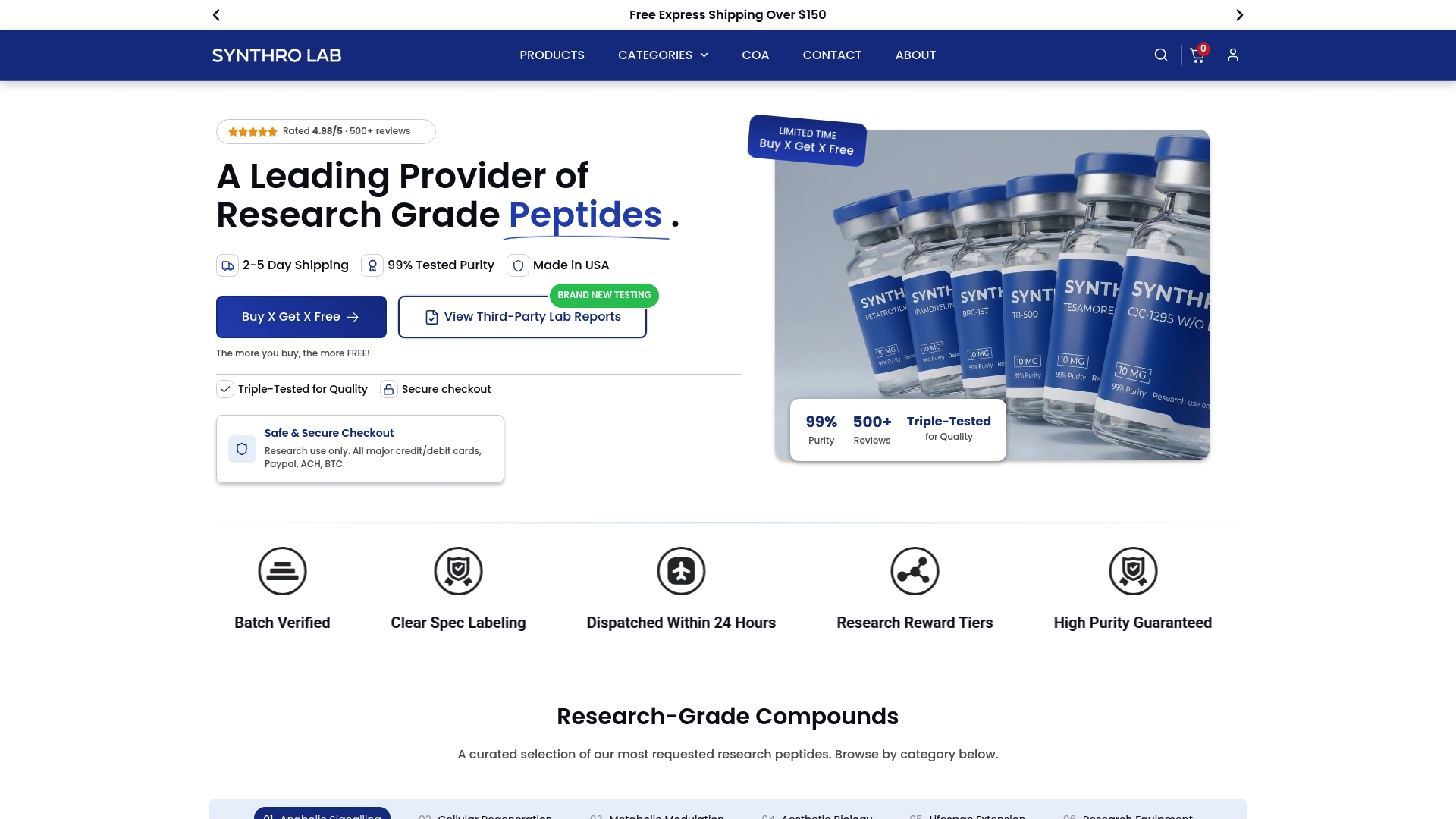
For researchers designing LL-37 experiments, Synthrolab also provides endotoxin-free reconstitution solutions and low-bind lab consumables to support the full experimental workflow. All materials are supplied for laboratory research use only and are not intended for human consumption. The peptide quality and CoA guide covers what to look for in a supplier CoA and how to evaluate purity claims before ordering.
Researchers with assay-specific QC questions, including endotoxin thresholds for particular cell systems or fragment selection guidance, can contact Synthrolab’s technical support team directly through the website.
Sources
This article is general information, not a substitute for advice from a qualified doctor. Consult a qualified healthcare professional about your own circumstances before acting on anything here.
Recommended
 21+ verification