Exosomes are nanosized, cell-derived extracellular vesicles that actively coordinate inflammation resolution, angiogenesis, and ECM remodeling, making them the leading cell-free modality in regenerative repair research today. A systematic rodent review found peak dermal therapeutic efficacy approximately 7 days after administration, with an odds ratio of 1.82 (95% CI [0.69, 2.95]), a figure that carries real weight when designing dosing schedules. Researchers working in this space should align isolation and characterization reporting with ISEV/MISEV standards from the outset, and Synthrolab provides research-grade compounds and cellular regeneration reagents for investigators building exosome-adjacent experimental programs.
The translational picture is this: preclinical evidence is strong across skin, nerve, tendon, cardiac, renal, and hepatic tissues. Early-phase clinical trials exist, but batch variability, inconsistent potency assays, and nomenclature ambiguity remain the primary barriers between a promising rodent result and a reproducible human study.
Key facts to anchor your reading:
- Exosomes range from 30–150 nm in diameter and are distinguished from larger microvesicles by their endosomal origin.
- Canonical surface markers CD9, CD63, and CD81 are the minimum reporting standard under MISEV guidelines.
- Stem cell-derived exosomes deliver miRNAs, VEGF, HGF, and TSG-6 to fibroblasts, endothelial cells, and macrophages, driving the core repair cascade.
- Engineered exosomes (eExo) consistently outperform naive preparations in targeted repair endpoints.
- FDA reviewers classify EV-based therapeutics as biologics, requiring full CMC documentation and potency metrics before IND submission.
Table of Contents
What is the role of exosomes in repair biology?
Exosomes belong to the broader family of extracellular vesicles (EVs), but their endosomal origin sets them apart. They form inside multivesicular bodies (MVBs) within the cell, then release into the extracellular space when MVBs fuse with the plasma membrane. That intracellular origin matters because it means exosomal cargo is not random membrane debris; it reflects the transcriptional and metabolic state of the parent cell at the moment of release.
Size is the first distinguishing feature: exosomes measure 30–150 nm, smaller than apoptotic bodies and most microvesicles. Under transmission electron microscopy, they appear as cup-shaped or spherical structures with a lipid bilayer. The canonical surface markers reported in MISEV-aligned studies are CD9, CD63, and CD81, tetraspanins that are enriched on the exosomal membrane and serve as identity markers in flow cytometry and Western blot characterization.
| Feature |
Exosomes |
Microvesicles |
Apoptotic Bodies |
| Diameter |
30–150 nm |
— |
— |
| Origin |
Endosomal (MVB) |
Plasma membrane budding |
Cell death |
| Canonical markers |
CD9, CD63, CD81 |
Integrins, selectins |
Phosphatidylserine |
| Cargo |
miRNA, mRNA, proteins, lipids |
Cytoplasmic proteins, DNA |
Organelles, nuclear fragments |
| Repair relevance |
Primary paracrine signaling |
Secondary signaling |
Clearance, phagocytosis |
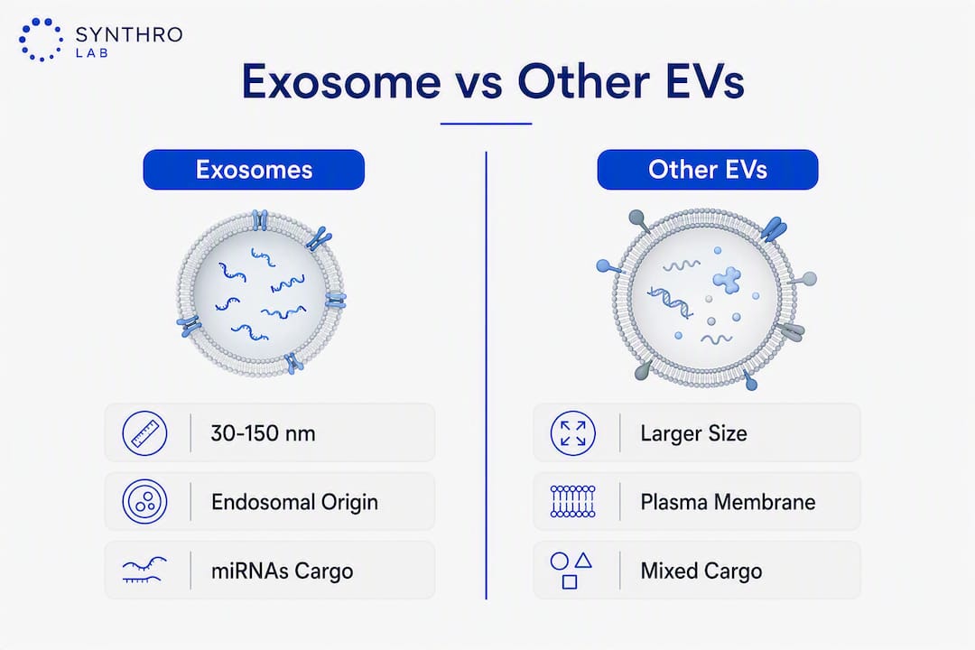
Cargo compartments span three categories. Small non-coding RNAs, particularly miRNAs, are the most studied; they enter recipient cells and suppress or activate gene networks post-transcriptionally. Proteins include growth factors, cytokines, and enzymes. Lipids, including sphingomyelin and cholesterol, contribute to membrane stability and receptor interactions.
Uptake by recipient cells occurs through three main routes: clathrin-mediated endocytosis, direct membrane fusion, and receptor-ligand interactions that trigger macropinocytosis. The route determines where cargo is delivered inside the cell and how quickly it acts. Membrane fusion deposits cargo directly into the cytoplasm; endocytosis routes it through endosomal compartments, which can degrade some RNA species before they reach their targets.
The repair-relevant cell types that respond to exosomal signals include:
- Fibroblasts: respond to exosomal TGF-β modulators and miRNAs that regulate collagen synthesis and myofibroblast differentiation.
- Endothelial cells: respond to VEGF and HIF-1α-related cargo that drives tube formation and neovascularization.
- Macrophages: shift from pro-inflammatory M1 to anti-inflammatory M2 phenotype in response to exosomal miR-146a and TSG-6.
- Schwann cells: receive signals that promote axonal regrowth and remyelination in peripheral nerve injury models.
- Keratinocytes: migrate and proliferate in response to exosomal growth factors during epidermal closure.
The conceptual flow is straightforward: a stressed or activated donor cell (say, a bone marrow MSC under hypoxic preconditioning) packages repair-relevant cargo into MVBs, releases exosomes into the local milieu, and those vesicles travel to fibroblasts, macrophages, or endothelial cells nearby, reprogramming their behavior without any direct cell-to-cell contact. That paracrine logic is what makes exosomes attractive as a cell-free therapeutic: you get the signaling benefit of a stem cell without transplanting a live cell.
Exosome biogenesis begins when the endosomal membrane invaginates inward, forming intraluminal vesicles (ILVs) inside the MVB. Two parallel sorting mechanisms determine which molecules end up inside those ILVs. The ESCRT (Endosomal Sorting Complexes Required for Transport) pathway recognizes ubiquitinated cargo proteins and loads them into ILVs through a stepwise ESCRT-0, -I, -II, -III assembly. ESCRT-independent pathways rely on ceramide-rich lipid microdomains and tetraspanin scaffolds (CD9, CD63) to cluster cargo without ubiquitin tagging.

Why does this matter experimentally? Because ESCRT-dependent and ESCRT-independent routes load different cargo profiles. Inhibiting one pathway shifts the molecular composition of the vesicles you collect, which changes their downstream repair activity. Researchers who treat “exosomes” as a homogenous reagent and ignore upstream biogenesis variables will see unexplained batch-to-batch variance in functional assays.
Once MVBs mature, they either fuse with lysosomes for degradation or traffic to the plasma membrane for release. The balance between those two fates is regulated by Rab GTPases (Rab27a, Rab27b, Rab35) and can be shifted pharmacologically, a lever that some engineering strategies exploit to increase exosome yield.
Cargo classes and their repair-relevant effects
| Cargo molecule |
Class |
Reported repair-relevant effect |
| miR-21 |
miRNA |
Suppresses PTEN; promotes fibroblast proliferation and angiogenesis |
| miR-146a |
miRNA |
Inhibits NF-κB; shifts macrophages toward M2; reduces IL-1β, IL-6 |
| miR-29 family |
miRNA |
Suppresses TGF-β/SMAD-driven fibrosis; reduces excess collagen deposition |
| HIF-1α-related cargo |
Protein/mRNA |
Upregulates VEGF expression; drives hypoxia-adaptive angiogenesis |
| TSG-6 |
Protein |
Anti-inflammatory; inhibits NF-κB; reduces neutrophil infiltration |
| VEGF |
Protein |
Direct angiogenic stimulus; promotes endothelial tube formation |
| HGF |
Protein |
Promotes hepatocyte and epithelial cell survival; anti-fibrotic |
| lncRNA MALAT1 |
lncRNA |
Modulates endothelial cell proliferation and migration |
The cargo profile is not fixed. Parent-cell state at the time of exosome collection is the dominant variable. Hypoxic preconditioning of MSCs, for example, increases HIF-1α-related cargo and VEGF loading, producing vesicles with stronger angiogenic activity than those collected from normoxic cultures. Inflammatory priming with LPS shifts the profile toward immunomodulatory miRNAs. This means cell-source control and preconditioning conditions are not secondary details in a methods section; they determine what you are actually testing.
Lipid cargo, though less studied than RNA and protein, contributes to membrane-mediated signaling. Lysophosphatidic acid and sphingosine-1-phosphate carried in exosomal membranes activate receptor pathways in fibroblasts and endothelial cells that parallel growth-factor signaling. Researchers designing lipidomic cargo profiles for repair applications are still in early territory, but the mechanistic rationale is solid.
How exosomes act across the phases of wound repair
Wound healing is conventionally divided into four overlapping phases: hemostasis, inflammation, proliferation, and remodeling. Exosomes do not act uniformly across all four; their effects are phase-specific, and the timing of administration relative to injury stage shapes the outcome.
Hemostasis. Platelet-derived EVs, including exosome-sized vesicles released during platelet activation, carry tissue factor and phosphatidylserine that accelerate thrombin generation and clot formation. This is the earliest EV-mediated contribution to repair, and it is largely endogenous rather than therapeutic. The clinical implication is that platelet-rich plasma (PRP)-derived EV preparations may carry procoagulant activity that needs to be characterized before systemic administration.
Inflammation resolution. The transition from M1 to M2 macrophage phenotype is one of the most consistently reported effects of therapeutic exosomes in repair models. MSC-derived exosomes suppress NF-κB, IL-1β, IL-6, and COX-2, reducing the proinflammatory burden that delays healing. miR-146a is a primary mediator: it targets IRAK1 and TRAF6, two upstream activators of NF-κB, and its delivery via exosomes produces a durable shift in macrophage gene expression rather than a transient cytokine block.
Proliferation and angiogenesis. Stem cell-derived exosomes carry VEGF, HGF, and HIF-1α-related cargo that directly stimulates endothelial tube formation and fibroblast migration. VEGF delivered via exosomal membrane-associated or luminal protein activates VEGFR2 on endothelial cells, triggering PI3K/Akt and MAPK/ERK pathways that drive neovascularization. In parallel, exosomal miR-21 suppresses PTEN in fibroblasts, increasing their proliferative response to growth factor signals. Keratinocyte migration, the rate-limiting step in epidermal closure, is accelerated by exosomal EGF-family proteins and miRNAs that reduce E-cadherin expression at the wound edge.

ECM remodeling. The MMP/TIMP balance governs whether a healing wound resolves cleanly or progresses to fibrosis. Exosomes from MSCs and ADSCs shift this balance by delivering miR-29 family members that suppress TGF-β/SMAD-driven collagen overproduction, and by modulating MMP-1, MMP-3, and TIMP-1 expression in fibroblasts. The net result in preclinical models is reduced scar area, improved tensile strength, and a collagen fiber architecture that more closely resembles uninjured tissue. HGF-carrying exosomes add an anti-fibrotic layer by competing with TGF-β1 signaling at the SMAD2/3 level.
The clinical relevance of these mechanisms is visible in preclinical outcomes: reduced scar formation in excisional wound models, improved nerve conduction velocity in crush injury models, and increased ejection fraction in myocardial infarction models. Each of those endpoints maps back to a specific pathway axis described above.
Which exosome sources work best for repair research?
The parent cell is the single biggest determinant of exosome therapeutic bias. Choosing a source without considering its regenerative profile is one of the most common design errors in translational exosome research.
Mesenchymal stem cell (MSC) sources — general
MSC-derived exosomes are the most studied class. They carry a broad immunomodulatory and pro-regenerative cargo profile and show applicability across neural, cardiovascular, hepatic, renal, articular, and oral tissues. The main limitation is donor variability: MSC passage number, tissue origin, and culture conditions all shift the cargo profile. Allogeneic use is feasible given low MHC-II expression on exosomes, but immunogenicity screening is still recommended.
Adipose-derived stem cells (ADSCs)
ADSC exosomes carry miR-132, miR-146a, and miR-125a, giving them a strong angiogenic and anti-fibrotic bias. They perform well in diabetic wound models where vascular insufficiency is a primary barrier. Adipose tissue is abundant and accessible, making autologous sourcing practical. The trade-off is that adipose tissue quality varies with donor metabolic status, which can affect exosome potency.
Bone marrow MSCs (BM-MSCs)
BM-MSC exosomes lean immunomodulatory rather than angiogenic. They are particularly well-studied in bone regeneration, cartilage repair, and hematologic recovery contexts. Harvest is more invasive than adipose, limiting autologous scalability, but allogeneic BM-MSC exosome banks are a realistic manufacturing path.
Umbilical cord / placental MSCs
These sources offer high proliferative capacity and consistent cargo profiles because donors are young and metabolically uniform. They are allogeneic by definition, but placental-derived exosomes show low immunogenicity in preclinical models. Regulatory classification of placental-derived products varies by jurisdiction and requires early FDA engagement in the US.
Platelet-derived EVs
Platelet-rich plasma-derived EVs are already in clinical use adjacent to exosome therapy (PRP injections). They carry PDGF, TGF-β1, and EGF, making them relevant for musculoskeletal and dermal repair. The challenge is that PRP preparations are highly heterogeneous and rarely meet MISEV characterization standards, making direct comparison with MSC exosome literature difficult.
Tissue-resident sources (tenocytes, keratinocytes, neurons)
Tissue-specific exosomes carry cargo optimized for their native environment. Tenocyte-derived exosomes, for example, carry tendon-specific matrix proteins and miRNAs that BM-MSC exosomes do not. The limitation is scalability: primary tenocytes are difficult to expand, and their exosome yield per culture volume is low compared to MSC lines.
Practical selection criteria to apply:
- Match source regenerative bias to the target tissue and primary repair deficit (angiogenesis vs. immunomodulation vs. matrix remodeling).
- Standardize passage number and culture conditions before comparing batches; collect exosomes at the same confluence and serum-starvation window.
- Document preconditioning conditions (normoxia vs. hypoxia, cytokine priming) in every methods section.
- For allogeneic preparations, include MHC profiling and immunogenicity screening in the preclinical package.
How should you isolate and characterize exosomes for repair studies?
Isolation method choice is not a minor technical detail. It determines purity, yield, and the degree of co-isolation of non-vesicular contaminants, all of which affect downstream functional assays and reproducibility.
| Isolation method |
Yield |
Purity |
Key limitation |
| Differential ultracentrifugation (dUC) |
High |
Moderate |
Co-pellets protein aggregates; time-intensive |
| Density gradient (iodixanol) |
Moderate |
High |
Low throughput; requires ultracentrifuge |
| Size-exclusion chromatography (SEC) |
Moderate |
High |
Dilutes sample; requires concentration step |
| Tangential flow filtration (TFF) |
High |
Moderate |
Retains large proteins; scale-dependent |
| Precipitation (PEG-based) |
High |
Low |
High contaminant co-precipitation; not MISEV-preferred |
Differential ultracentrifugation remains the most widely used method, but ISEV/MISEV guidelines consistently note that it co-pellets protein aggregates and HDL particles, inflating particle counts and distorting cargo profiles. For translational studies where potency and cargo identity matter, SEC or density gradient methods are preferred despite lower throughput.
Minimum characterization to report (MISEV-aligned)
Every repair-focused exosome study should report:
- Particle size distribution: nanoparticle tracking analysis (NTA) or dynamic light scattering (DLS), with mean and mode diameter.
- Morphology: transmission electron microscopy (TEM) confirming cup-shaped vesicle morphology.
- Tetraspanin markers: Western blot or flow cytometry for CD9, CD63, and CD81 (positive enrichment).
- Contamination markers: absence or depletion of calnexin (ER marker), GM130 (Golgi), and albumin (serum contamination).
- Particle-to-protein ratio: a key purity metric; ratios below 1×10¹⁰ particles/µg protein suggest significant protein co-contamination.
- RNA/protein cargo profiling: at minimum, a representative miRNA panel and total protein quantification.
- Sterility and endotoxin: LAL assay for endotoxin; sterility testing for any preparation intended for in vivo use.
- Batch certificate of analysis (COA): particle count, size, marker expression, and endotoxin result per batch.
Potency assays tied to repair endpoints are the most underdeveloped area in the field. A particle count tells you how much you have; it does not tell you whether it works. Functional potency assays to consider include scratch-wound migration assays (fibroblast or keratinocyte), tube formation assays (HUVEC), macrophage polarization assays (M1→M2 shift by flow cytometry), and collagen gel contraction assays. Map the assay to the primary mechanism you are claiming.
Pro Tip: When reporting isolation methods in a manuscript or regulatory submission, include the particle-to-protein ratio alongside particle count and size. Reviewers and FDA CMC teams increasingly treat this ratio as a minimum purity indicator, and omitting it flags the preparation as inadequately characterized.
What delivery routes and timing work best for exosome repair studies?
Delivery strategy is where many preclinical exosome studies lose translational relevance. A preparation that works beautifully when injected directly into a wound edge in a mouse may fail systemically in a larger model, not because the biology is wrong, but because the pharmacokinetics are entirely different.
Local injection is the most direct route for wound, tendon, and nerve repair applications. It maximizes local concentration, reduces systemic clearance, and allows dose titration against a defined tissue volume. The limitation is that a single bolus injection clears rapidly; exosomes injected into soft tissue have a half-life measured in hours without a retention matrix.
Topical and hydrogel delivery addresses the clearance problem for dermal applications. Incorporating exosomes into fibrin, hyaluronic acid, or collagen hydrogels extends local residence time and provides a sustained-release profile that better matches the multi-day repair window. This approach is particularly well-supported for cutaneous wound healing, where the peak therapeutic efficacy at approximately 7 days post-administration (odds ratio 1.82 in the rodent systematic review) suggests that sustained local exposure through the proliferative phase is more important than a high initial bolus.
Scaffold incorporation extends the hydrogel concept to three-dimensional biomaterial constructs for bone, cartilage, and tendon repair. Electrospun nanofiber scaffolds, decellularized ECM, and 3D-printed constructs have all been used as exosome carriers in preclinical models. The advantage is spatial control: exosomes are released where the scaffold is, not distributed systemically.
Systemic intravenous (IV) delivery is the least efficient route for tissue-specific repair. After IV injection, exosomes distribute rapidly to liver, spleen, and lung, with minimal accumulation at peripheral injury sites unless surface-modified with targeting ligands. Biodistribution studies using DiI or DiR-labeled exosomes consistently show hepatic and splenic accumulation within 24 hours, with signal at injury sites only when local inflammation creates a homing gradient.
Key timing and dosing considerations:
- The 7-day peak efficacy window in dermal models implies that dosing schedules should include at least one administration within the first week, with readouts at days 7, 14, and 28.
- Repeat dosing (days 1, 3, 7) outperforms single-dose protocols in several tendon and nerve models, though the optimal interval is tissue-specific.
- Dose-ranging studies are mandatory before efficacy claims; most published rodent studies use 50–200 µg total protein equivalent per dose, but this metric is only meaningful if the particle-to-protein ratio is also reported.
- Pharmacokinetic studies should include at least two time points (24 hours and 72 hours post-injection) and use orthogonal detection methods (imaging plus tissue extraction).
Pro Tip: For any repair indication where the target tissue is accessible (skin, tendon, joint), favor a biomaterial-embedded local delivery approach over systemic IV in early preclinical work. You will get cleaner dose-response data, better retention evidence, and a more defensible translational rationale for the IND package.
What does the preclinical and clinical evidence show across tissues?
The evidence base is not uniform. Cutaneous wound healing and peripheral nerve regeneration have the deepest preclinical literature; cardiac and renal applications are earlier-stage with more variable results.
| Tissue / indication |
Key preclinical outcomes |
Clinical trial status |
| Cutaneous wound healing |
Accelerated closure, reduced scar, increased angiogenesis, M2 polarization |
Multiple early-phase trials registered; ADSC and MSC exosome preparations most common |
| Peripheral nerve regeneration |
Improved axonal regrowth, increased myelin thickness, better conduction velocity |
Phase I/II trials in progress; limited published results |
| Tendon / ligament repair |
Reduced NF-κB/IL-1β, improved COL1A1/COL3A1 ratio, better tensile strength |
Mostly preclinical; a small number of early trials registered |
| Skeletal muscle repair |
Reduced fibrosis, improved fiber regeneration, macrophage polarization shift |
Preclinical; no large trials published |
| Cardiac (post-MI) |
Improved ejection fraction, reduced infarct size, angiogenesis |
Phase I trials exist; results heterogeneous |
| Bone / cartilage |
Increased osteogenesis/chondrogenesis markers, reduced inflammation |
Early-phase trials; mostly in vitro/small animal |
| Renal injury |
Reduced tubular apoptosis, improved creatinine in AKI models |
Preclinical; very limited clinical data |
| Hepatic injury / fibrosis |
Anti-fibrotic, reduced ALT/AST, improved hepatocyte survival |
Preclinical; early clinical interest |
Exosomes facilitate tendon, skeletal muscle, and peripheral nerve regeneration primarily through macrophage polarization, NF-κB suppression, and ECM gene rebalancing (COL1A1, COL3A1, MMP/TIMP). In nerve models, Schwann cell activation and axonal guidance are the additional mechanisms that distinguish neural repair from soft-tissue repair.
Cardiac applications show the most heterogeneous clinical results. Preclinical MI models consistently show improved ejection fraction and reduced infarct size with MSC-derived exosomes, but the translation to human trials has been complicated by delivery timing (acute vs. subacute administration), route (intracoronary vs. IV), and product variability. Trials registered on ClinicalTrials.gov include early-phase studies in chronic wound healing and GvHD, where MSC exosome immunomodulatory properties are the primary mechanism of interest.
Limitations in the current clinical literature are significant. Most trials enroll fewer than 30 participants, use heterogeneous product definitions (some report particle count, others report protein equivalent, many report neither), and lack standardized potency readouts. Without a shared functional potency assay, comparing results across trials is essentially impossible. This is not a minor methodological complaint; it is the reason the field has not yet produced a Phase III trial with a clear efficacy signal.
What safety concerns and regulatory barriers should researchers know?
Safety concerns in exosome therapeutics are real but manageable with systematic preclinical screening. The risks are not hypothetical; they follow directly from the biology.
Pro-tumorigenic cargo risk. Exosomes from cancer cells carry oncogenic miRNAs and proteins that promote tumor progression. The concern for therapeutic preparations is whether exosomes from non-malignant parent cells carry low-level pro-proliferative signals that could accelerate occult malignancy in immunocompromised recipients. Preclinical genotoxicity and tumorigenicity screens are mandatory for any preparation intended for human use.
Immune activation. While MSC-derived exosomes generally show low immunogenicity, they carry MHC-I and can present peptide antigens. Repeated dosing in allogeneic settings may generate adaptive immune responses. Complement activation by exosomal phosphatidylserine is a separate concern for IV-administered preparations.
Infectious agent transmission. Exosomes derived from virus-infected cells can carry viral RNA or proteins. Donor cell screening for HIV, HBV, HCV, and CMV is a minimum requirement. For allogeneic preparations, this mirrors blood product safety standards.
Pro-fibrotic signals. TGF-β1 is present in many MSC exosome preparations. At high doses or in fibrosis-prone tissues, exosomal TGF-β1 could drive rather than resolve scarring. Dose-ranging studies should include fibrosis markers (hydroxyproline content, α-SMA expression) as safety endpoints, not just efficacy endpoints.
Translational barriers beyond safety:
- Manufacturing scale: most academic exosome preparations use 2D cell culture, which cannot produce the quantities needed for clinical trials. Bioreactor-based 3D culture and hollow-fiber systems are being developed but are not yet standardized.
- Batch variability: cargo profiles shift with passage number, serum lot, and collection timing. Without a validated potency assay, batch release is based on physical characterization alone, which is insufficient.
- Nomenclature ambiguity: the term “exosome” is used inconsistently in the literature. Many published studies isolate a mixed EV population and call it exosomes. This creates a reproducibility problem when other groups try to replicate results.
US regulatory framework (FDA expectations for EV-based biologics):
- EV therapeutics are classified as biological products under 21 CFR Part 600 and require a Biologics License Application (BLA) or IND for clinical investigation.
- CMC documentation must include: cell banking, production process description, release testing (particle count, size, sterility, endotoxin, identity markers), and stability data.
- Potency assay: FDA expects a mechanism-relevant functional assay, not just physical characterization. The assay must be qualified before Phase II.
- Preclinical package for IND: pharmacology/efficacy data in at least one relevant animal model, toxicology (repeat-dose, biodistribution), and genotoxicity if the preparation contains nucleic acid cargo.
- Combination products (exosomes in a scaffold or hydrogel) may require dual regulatory pathway coordination between CBER and CDRH.
This article provides general scientific and regulatory information, not legal or clinical advice. Researchers should confirm current FDA guidance with a qualified regulatory professional or directly with the relevant FDA center before initiating IND-enabling studies.
How do engineered exosomes improve on native preparations?
Native exosomes from unstimulated parent cells often carry insufficient cargo concentration for high-demand repair applications. The field has moved decisively toward engineered exosomes (eExo) as the necessary next step, and the evidence supports that shift.
Engineering strategies fall into three categories:
Pre-exosomal (parent cell modification)
- Hypoxic preconditioning (1–5% O₂) increases HIF-1α-driven cargo loading and VEGF content, producing exosomes with stronger angiogenic activity.
- Cytokine priming (IFN-γ, TNF-α) shifts the immunomodulatory cargo profile toward anti-inflammatory mediators.
- Genetic overexpression of specific miRNAs (miR-21, miR-146a) in parent cells produces exosomes enriched for those sequences.
Post-exosomal (direct cargo loading)
- Electroporation is the most widely used method for loading exogenous siRNA, miRNA mimics, or mRNA into isolated exosomes. Platforms including EXPLOR and co-incubation methods have been used to load therapeutic RNAs and proteins, with improved wound-healing outcomes in animal models.
- Sonication and saponin-assisted loading are alternatives with different cargo size tolerances.
- Surface modification with targeting peptides (RGD, CXCR4 ligands) improves tissue-specific homing after systemic administration.
Biohybrid approaches
- Incorporating exosomes into hydrogels, nanofiber scaffolds, or liposome-exosome hybrids extends release kinetics and adds structural support for tissue defects.
- Exosome-coated nanoparticles combine the targeting capacity of synthetic nanocarriers with the biocompatibility of exosomal membranes.
| Approach |
Primary benefit |
Key limitation |
| Hypoxic preconditioning |
Increased VEGF/HIF-1α cargo; higher yield |
Requires controlled hypoxia chamber; adds process complexity |
| Genetic cargo loading (overexpression) |
Consistent, high-level target miRNA enrichment |
Requires stable transfection; regulatory complexity for GMO-derived products |
| Electroporation loading |
Flexible; works with diverse cargo types |
Can damage vesicle membrane; loading efficiency varies |
| Surface peptide modification |
Tissue-specific targeting after systemic delivery |
Peptide stability in vivo; manufacturing reproducibility |
| Biohybrid scaffold |
Sustained local release; structural support |
Scaffold biocompatibility must be independently validated |
eExo strategies are viewed as necessary for high-demand tissue regeneration where native exosome concentrations are insufficient. The translational checklist for moving an eExo concept toward IND-enabling studies should include:
- Dose-ranging with the engineered preparation (not the native exosome dose as a proxy).
- Stability data for the modified cargo under storage and physiological conditions.
- Targeted biodistribution study confirming that surface modifications actually improve tissue accumulation.
- Scalable isolation method validated for the engineered preparation (electroporation efficiency must be confirmed at scale).
- Potency readout tied directly to the engineered mechanism (e.g., if you loaded miR-146a, the potency assay should measure NF-κB suppression or M2 polarization, not just particle count).
For manufacturing scale-up, bioreactor-based 3D culture (hollow-fiber, stirred-tank) is the only realistic path to clinical quantities. Yield from 2D flasks is inadequate for multi-dose clinical protocols, and the process change from 2D to 3D must be validated with a comparability study showing equivalent cargo profiles and potency.
Best practices for designing reproducible exosome repair studies
The reproducibility problem in exosome research is not primarily a statistics problem. It is an upstream variable-control problem. Studies that treat “exosomes” as a single reagent without specifying source, passage, isolation method, characterization, and potency will produce results that cannot be replicated, regardless of sample size.
Here is a practical checklist for designing a repair-focused exosome study that will survive peer review and, if relevant, regulatory scrutiny:
- Define and document the parent cell source: species, tissue origin, passage number, culture medium (including serum lot), and any preconditioning conditions. Report these in the methods, not just the supplementary materials.
- Select and validate an isolation method: choose SEC or density gradient for high-purity applications; use dUC only if throughput is the primary constraint and you report the particle-to-protein ratio. Never use precipitation-only methods for translational studies.
- Report MISEV-minimum characterization: NTA (size and concentration), TEM (morphology), Western blot for CD9/CD63/CD81 (positive) and calnexin/GM130 (negative), and endotoxin result. This is the floor, not the ceiling.
- Include a mechanism-relevant potency assay: scratch-wound migration, tube formation, or macrophage polarization assay run in parallel with each batch. Batch release should be conditional on passing the potency threshold.
- Use a standardized in vivo model with defined readout timepoints: for dermal repair, the recommended schedule is days 3, 7, 14, and 28 post-injury. For nerve repair, add electrophysiology at 4 and 8 weeks. For tendon, biomechanical testing at 4 and 8 weeks. Align with the tissue repair research protocol framework for endpoint selection.
- Power the study appropriately: most published exosome studies are underpowered. Use a pilot study or published variance estimates to calculate sample size before committing to a full experiment. Report the power calculation in the methods.
- Blind outcome assessment: histology scoring, functional assays, and image analysis should be performed by an observer blinded to treatment group. This is non-negotiable for any study intended for publication in a high-impact journal or regulatory submission.
- Include appropriate controls: vehicle control (the carrier without exosomes), positive control (a validated repair stimulus), and, where possible, a heat-inactivated exosome control to distinguish vesicle-mediated from soluble-factor-mediated effects.
- Report raw cargo profiles: miRNA profiling (small RNA-seq or qPCR panel) and total protein quantification per batch. Deposit raw data in a public repository (GEO, Zenodo) to enable meta-analysis.
- Provide a batch COA: particle count, size distribution, marker expression, endotoxin, and sterility result for every batch used in the study. This is standard practice for any research-grade biological reagent and is the minimum expectation for regulatory submissions.
A sample experimental timeline for a rodent dermal repair study: administer exosomes at day 0 (injury) and day 3; collect tissue at days 7, 14, and 28; run histology (H&E, Masson’s trichrome), immunohistochemistry (CD31 for angiogenesis, CD68/CD206 for macrophage phenotype, α-SMA for myofibroblasts), and biomechanical testing at day 28. Potency assay on the administered batch should be completed before the in vivo experiment starts, not after.
For researchers building out their experimental toolkit, Synthrolab’s research-grade peptide and compound catalog includes batch-tested reagents with certificates of analysis, which can serve as complementary tools in repair pathway studies alongside exosome preparations.
Key Takeaways
Exosomes coordinate repair through paracrine cargo delivery, with peak dermal efficacy at approximately 7 days post-administration, and translational success depends on standardized isolation, mechanism-relevant potency assays, and engineered cargo strategies.
| Point |
Details |
| Peak dermal efficacy timing |
Systematic rodent review found maximum therapeutic effect at approximately 7 days post-administration (odds ratio 1.82). |
| MISEV characterization is the floor |
Every repair study must report size, TEM morphology, CD9/CD63/CD81, contamination markers, and particle-to-protein ratio. |
| Source determines therapeutic bias |
ADSC exosomes favor angiogenesis and anti-fibrosis; BM-MSC exosomes favor immunomodulation; match source to repair deficit. |
| eExo outperform native preparations |
Preconditioning, cargo loading, and biohybrid approaches are necessary for high-demand regeneration targets. |
| Potency assay is mandatory |
Particle count alone is insufficient; functional assays (scratch migration, tube formation, M2 polarization) must gate batch release. |
The field is moving faster than the standards can keep up
The mechanistic case for exosomes in repair is genuinely strong. The cargo biology is well-characterized, the pathway evidence is consistent across multiple tissue types, and the preclinical results are reproducible when upstream variables are controlled. What is not strong is the translational infrastructure around it.
Here is what most reviews understate: the reproducibility crisis in exosome research is not a problem of bad science. It is a problem of treating a heterogeneous biological preparation as if it were a defined chemical entity. A “50 µg exosome dose” from a passage-8 BM-MSC culture under normoxic conditions is a fundamentally different reagent than a “50 µg exosome dose” from a passage-3 ADSC culture under hypoxic preconditioning, even if both pass the same NTA and Western blot criteria. The field has been slow to accept that cargo profiling is not optional; it is the identity test.
The shift toward eExo is the right direction, but it introduces a new problem: regulatory complexity. Genetically modified parent cells, electroporated exosomes carrying synthetic miRNA mimics, and biohybrid scaffold constructs each sit in a different regulatory category. Researchers who design elegant eExo systems without early FDA engagement are building toward an IND package that will require significant rework. The time to think about CMC documentation is at the experimental design stage, not after you have generated two years of efficacy data.
What should researchers prioritize? Three things, in order. First, fix the upstream: standardize your source, passage, and preconditioning before you run a single in vivo experiment. Second, build a potency assay that is mechanistically linked to your repair claim and use it as a batch release gate, not a post-hoc characterization. Third, if your target indication requires high-dose or repeat-dose administration, start the eExo engineering work now, because native exosome concentrations from 2D culture will not get you to clinical quantities or clinical potency.
The biology is ready. The infrastructure is catching up. Researchers who build the standardization into their workflow from the start will be the ones with translatable data when the clinical window opens.
Synthrolab supplies research-grade peptides and laboratory compounds, each independently batch-tested with a full certificate of analysis, for investigators running cellular signaling, recovery pathway, and regenerative mechanism studies. Researchers building exosome-adjacent experimental programs, or exploring complementary repair modalities such as GHK-Cu, BPC-157, and TB-500, will find Synthrolab’s catalog a practical starting point for research-grade reagents with documented purity.
Recommended
 21+ verification